4QC6

Crystal structure of aminoglycoside 6'-acetyltransferase-Ie


Experimental Data Snapshot

  • Method: X-RAY DIFFRACTION
  • Resolution: 1.30 Å
  • R-Value Free: 0.166 
  • R-Value Work: 0.147 
  • R-Value Observed: 0.148 

wwPDB Validation   3D Report Full Report


Ligand Structure Quality Assessment 


This is version 1.1 of the entry. See complete history


Literature

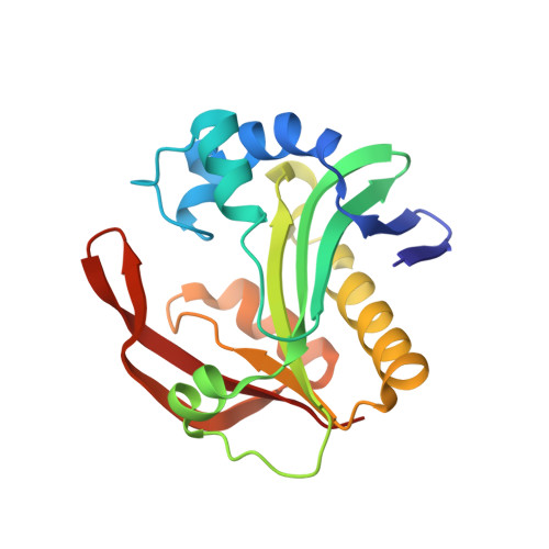
Structure of the bifunctional aminoglycoside-resistance enzyme AAC(6')-Ie-APH(2'')-Ia revealed by crystallographic and small-angle X-ray scattering analysis.

Smith, C.A.Toth, M.Weiss, T.M.Frase, H.Vakulenko, S.B.

(2014) Acta Crystallogr D Biol Crystallogr 70: 2754-2764

  • DOI: https://doi.org/10.1107/S1399004714017635
  • Primary Citation of Related Structures:  
    4QC6

  • PubMed Abstract: 

    Broad-spectrum resistance to aminoglycoside antibiotics in clinically important Gram-positive staphylococcal and enterococcal pathogens is primarily conferred by the bifunctional enzyme AAC(6')-Ie-APH(2'')-Ia. This enzyme possesses an N-terminal coenzyme A-dependent acetyltransferase domain [AAC(6')-Ie] and a C-terminal GTP-dependent phosphotransferase domain [APH(2'')-Ia], and together they produce resistance to almost all known aminoglycosides in clinical use. Despite considerable effort over the last two or more decades, structural details of AAC(6')-Ie-APH(2'')-Ia have remained elusive. In a recent breakthrough, the structure of the isolated C-terminal APH(2'')-Ia enzyme was determined as the binary Mg2GDP complex. Here, the high-resolution structure of the N-terminal AAC(6')-Ie enzyme is reported as a ternary kanamycin/coenzyme A abortive complex. The structure of the full-length bifunctional enzyme has subsequently been elucidated based upon small-angle X-ray scattering data using the two crystallographic models. The AAC(6')-Ie enzyme is joined to APH(2'')-Ia by a short, predominantly rigid linker at the N-terminal end of a long α-helix. This α-helix is in turn intrinsically associated with the N-terminus of APH(2'')-Ia. This structural arrangement supports earlier observations that the presence of the intact α-helix is essential to the activity of both functionalities of the full-length AAC(6')-Ie-APH(2'')-Ia enzyme.


  • Organizational Affiliation

    Stanford Synchrotron Radiation Lightsource, Stanford University, Menlo Park, CA 94025, USA.


Macromolecules
Find similar proteins by:  (by identity cutoff)  |  3D Structure
Entity ID: 1
MoleculeChains Sequence LengthOrganismDetailsImage
Bifunctional AAC/APH
A, B
179Staphylococcus warneriMutation(s): 0 
Gene Names: aacA-aphD
EC: 2.3.1 (PDB Primary Data), 2.7.1 (PDB Primary Data)
UniProt
Find proteins for Q7ATH7 (Staphylococcus warneri)
Explore Q7ATH7 
Go to UniProtKB:  Q7ATH7
Entity Groups  
Sequence Clusters30% Identity50% Identity70% Identity90% Identity95% Identity100% Identity
UniProt GroupQ7ATH7
Sequence Annotations
Expand
  • Reference Sequence
Small Molecules
Ligands 3 Unique
IDChains Name / Formula / InChI Key2D Diagram3D Interactions
30N
Query on 30N

Download Ideal Coordinates CCD File 
D [auth A],
H [auth B]
(3R,5S,9R)-1-[(2R,3S,4R,5R)-5-(6-amino-9H-purin-9-yl)-4-hydroxy-3-(phosphonooxy)tetrahydrofuran-2-yl]-3,5,9-trihydroxy-8,8-dimethyl-10,14-dioxo-2,4,6-trioxa-11,15-diaza-3,5-diphosphaheptadecane-17-sulfinic acid 3,5-dioxide (non-preferred name)
C21 H36 N7 O18 P3 S
HHNOQZRCHLIIMA-IBOSZNHHSA-N
KAN
Query on KAN

Download Ideal Coordinates CCD File 
C [auth A],
G [auth B]
KANAMYCIN A
C18 H36 N4 O11
SBUJHOSQTJFQJX-NOAMYHISSA-N
FMT
Query on FMT

Download Ideal Coordinates CCD File 
E [auth A],
F [auth A],
I [auth B],
J [auth B],
K [auth B]
FORMIC ACID
C H2 O2
BDAGIHXWWSANSR-UHFFFAOYSA-N
Experimental Data & Validation

Experimental Data

  • Method: X-RAY DIFFRACTION
  • Resolution: 1.30 Å
  • R-Value Free: 0.166 
  • R-Value Work: 0.147 
  • R-Value Observed: 0.148 
  • Space Group: P 21 21 21
Unit Cell:
Length ( Å )Angle ( ˚ )
a = 52.83α = 90
b = 89.72β = 90
c = 96.96γ = 90
Software Package:
Software NamePurpose
Blu-Icedata collection
MOLREPphasing
PHENIXrefinement
XDSdata reduction
SCALAdata scaling

Structure Validation

View Full Validation Report


Ligand Structure Quality Assessment 


Entry History 

Deposition Data

Revision History  (Full details and data files)

  • Version 1.0: 2014-10-22
    Type: Initial release
  • Version 1.1: 2024-02-28
    Changes: Data collection, Database references, Derived calculations