4PXO

Crystal structure of Maleylacetoacetate isomerase from Methylobacteriu extorquens AM1 WITH BOUND MALONATE AND GSH (TARGET EFI-507068)


Experimental Data Snapshot

  • Method: X-RAY DIFFRACTION
  • Resolution: 1.80 Å
  • R-Value Free: 0.174 
  • R-Value Work: 0.144 
  • R-Value Observed: 0.145 

wwPDB Validation   3D Report Full Report


Ligand Structure Quality Assessment 


This is version 1.3 of the entry. See complete history


Literature

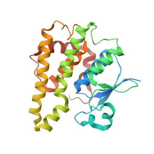
Crystal structure of glutathione s-transferase zeta from Methylobacterium extorquens (TARGET EFI-507068)

Patskovsky, Y.Toro, R.Bhosle, R.Hillerich, B.Seidel, R.D.Washington, E.Scott Glenn, A.Chowdhury, S.Evans, B.Hammonds, J.Imker, H.J.Al Obaidi, N.Stead, M.Love, J.Gerlt, J.A.Armstrong, R.N.Almo, S.C.Enzyme Function Initiative (EFI)

To be published.

Macromolecules
Find similar proteins by:  (by identity cutoff)  |  3D Structure
Entity ID: 1
MoleculeChains Sequence LengthOrganismDetailsImage
Maleylacetoacetate isomerase (Glutathione S-transferase)
A, B
238Methylorubrum extorquens AM1Mutation(s): 0 
Gene Names: maiAMexAM1_META1p2830
EC: 2.5.1.18 (PDB Primary Data), 5.2.1.2 (PDB Primary Data)
UniProt
Find proteins for C5ATQ9 (Methylorubrum extorquens (strain ATCC 14718 / DSM 1338 / JCM 2805 / NCIMB 9133 / AM1))
Explore C5ATQ9 
Go to UniProtKB:  C5ATQ9
Entity Groups  
Sequence Clusters30% Identity50% Identity70% Identity90% Identity95% Identity100% Identity
UniProt GroupC5ATQ9
Sequence Annotations
Expand
  • Reference Sequence
Small Molecules
Ligands 3 Unique
IDChains Name / Formula / InChI Key2D Diagram3D Interactions
GSH
Query on GSH

Download Ideal Coordinates CCD File 
D [auth A],
I [auth B]
GLUTATHIONE
C10 H17 N3 O6 S
RWSXRVCMGQZWBV-WDSKDSINSA-N
MLA
Query on MLA

Download Ideal Coordinates CCD File 
C [auth A],
H [auth B]
MALONIC ACID
C3 H4 O4
OFOBLEOULBTSOW-UHFFFAOYSA-N
EDO
Query on EDO

Download Ideal Coordinates CCD File 
E [auth A]
F [auth A]
G [auth A]
J [auth B]
K [auth B]
E [auth A],
F [auth A],
G [auth A],
J [auth B],
K [auth B],
L [auth B],
M [auth B],
N [auth B]
1,2-ETHANEDIOL
C2 H6 O2
LYCAIKOWRPUZTN-UHFFFAOYSA-N
Experimental Data & Validation

Experimental Data

  • Method: X-RAY DIFFRACTION
  • Resolution: 1.80 Å
  • R-Value Free: 0.174 
  • R-Value Work: 0.144 
  • R-Value Observed: 0.145 
  • Space Group: P 65 2 2
Unit Cell:
Length ( Å )Angle ( ˚ )
a = 109.628α = 90
b = 109.628β = 90
c = 185.367γ = 120
Software Package:
Software NamePurpose
PHASERphasing
REFMACrefinement
HKL-3000data reduction
HKL-3000data scaling

Structure Validation

View Full Validation Report


Ligand Structure Quality Assessment 


Entry History 

Revision History  (Full details and data files)

  • Version 1.0: 2014-04-09
    Type: Initial release
  • Version 1.1: 2015-04-29
    Changes: Non-polymer description
  • Version 1.2: 2018-01-24
    Changes: Database references, Structure summary
  • Version 1.3: 2023-09-20
    Changes: Data collection, Database references, Derived calculations, Refinement description