4PR7

KdgM porin in complex with disordered oligogalacturonate


Experimental Data Snapshot

  • Method: X-RAY DIFFRACTION
  • Resolution: 2.10 Å
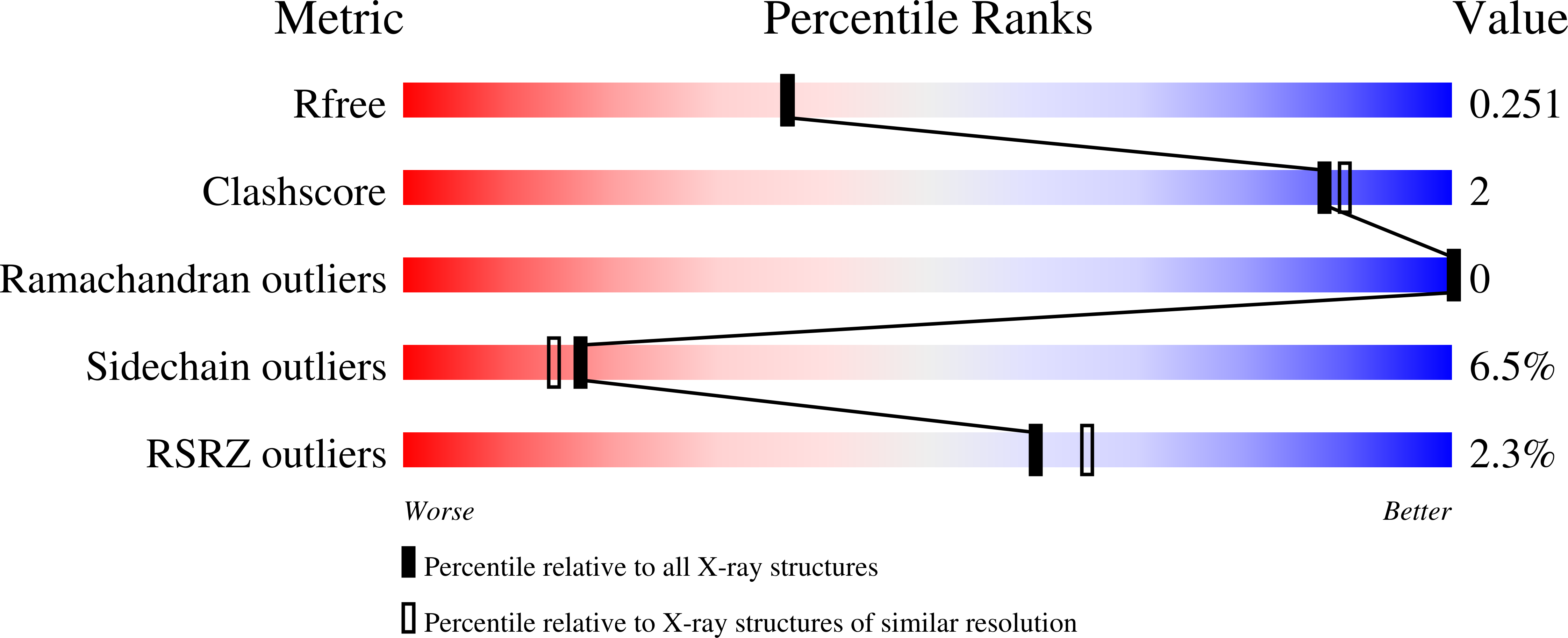
  • R-Value Free: 0.245 
  • R-Value Work: 0.208 
  • R-Value Observed: 0.210 

wwPDB Validation   3D Report Full Report


Ligand Structure Quality Assessment 


This is version 2.1 of the entry. See complete history


Literature

Structure of the oligogalacturonate-specific KdgM porin.

Hutter, C.A.Lehner, R.Wirth, C.h.Condemine, G.Peneff, C.Schirmer, T.

(2014) Acta Crystallogr D Biol Crystallogr 70: 1770-1778

  • DOI: https://doi.org/10.1107/S1399004714007147
  • Primary Citation of Related Structures:  
    4FQE, 4PR7

  • PubMed Abstract: 

    The phytopathogenic Gram-negative bacterium Dickeya dadantii (Erwinia chrysanthemi) feeds on plant cell walls by secreting pectinases and utilizing the oligogalacturanate products. An outer membrane porin, KdgM, is indispensable for the uptake of these acidic oligosaccharides. Here, the crystal structure of KdgM determined to 1.9 Å resolution is presented. KdgM is folded into a regular 12-stranded antiparallel β-barrel with a circular cross-section defining a transmembrane pore with a minimal radius of 3.1 Å. Most of the loops that would face the cell exterior in vivo are disordered, but nevertheless mediate contact between densely packed membrane-like layers in the crystal. The channel is lined by two tracks of arginine residues facing each other across the pore, a feature that is conserved within the KdgM family and is likely to facilitate the diffusion of acidic oligosaccharides.


  • Organizational Affiliation

    Focal Area of Structural Biology and Biophysics, Biozentrum, University of Basel, Klingelbergstrasse 70, CH-4056 Basel, Switzerland.


Macromolecules
Find similar proteins by:  (by identity cutoff)  |  3D Structure
Entity ID: 1
MoleculeChains Sequence LengthOrganismDetailsImage
Oligogalacturonate-specific porin KdgM222Dickeya dadantiiMutation(s): 0 
Gene Names: kdgMDda3937_03362
Membrane Entity: Yes 
UniProt
Find proteins for Q934G3 (Dickeya dadantii (strain 3937))
Explore Q934G3 
Go to UniProtKB:  Q934G3
Entity Groups  
Sequence Clusters30% Identity50% Identity70% Identity90% Identity95% Identity100% Identity
UniProt GroupQ934G3
Sequence Annotations
Expand
  • Reference Sequence
Oligosaccharides

Help

Entity ID: 2
MoleculeChains Length2D Diagram Glycosylation3D Interactions
alpha-D-galactopyranuronic acid-(1-4)-alpha-D-galactopyranuronic acid-(1-4)-alpha-D-galactopyranuronic acid-(1-4)-alpha-D-galactopyranuronic acid-(1-4)-alpha-D-galactopyranuronic acid
B
5N/A
Glycosylation Resources
GlyTouCan:  G48720UK
GlyCosmos:  G48720UK
Experimental Data & Validation

Experimental Data

  • Method: X-RAY DIFFRACTION
  • Resolution: 2.10 Å
  • R-Value Free: 0.245 
  • R-Value Work: 0.208 
  • R-Value Observed: 0.210 
  • Space Group: P 1 21 1
Unit Cell:
Length ( Å )Angle ( ˚ )
a = 36.8569α = 90
b = 59.7311β = 106.5412
c = 51.5959γ = 90
Software Package:
Software NamePurpose
REFMACrefinement
SCALAdata scaling
PDB_EXTRACTdata extraction
MOSFLMdata reduction
PHASERphasing

Structure Validation

View Full Validation Report


Ligand Structure Quality Assessment 


Entry History 

Deposition Data

Revision History  (Full details and data files)

  • Version 1.0: 2014-06-11
    Type: Initial release
  • Version 1.1: 2014-09-24
    Changes: Database references
  • Version 2.0: 2020-07-29
    Type: Remediation
    Reason: Carbohydrate remediation
    Changes: Advisory, Atomic model, Data collection, Database references, Derived calculations, Structure summary
  • Version 2.1: 2023-09-20
    Changes: Advisory, Data collection, Database references, Refinement description, Structure summary