4PPH

Crystal structure of conglutin gamma, a unique basic 7S globulin from lupine seeds


Experimental Data Snapshot

  • Method: X-RAY DIFFRACTION
  • Resolution: 2.01 Å
  • R-Value Free: 0.174 
  • R-Value Work: 0.145 
  • R-Value Observed: 0.146 

wwPDB Validation   3D Report Full Report


Ligand Structure Quality Assessment 


This is version 2.1 of the entry. See complete history


Literature

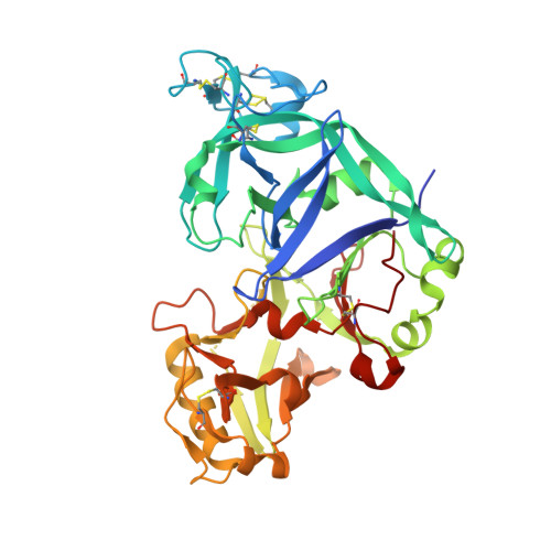
Structure of gamma-conglutin: insight into the quaternary structure of 7S basic globulins from legumes.

Czubinski, J.Barciszewski, J.Gilski, M.Szpotkowski, K.Debski, J.Lampart-Szczapa, E.Jaskolski, M.

(2015) Acta Crystallogr D Biol Crystallogr 71: 224-238

  • DOI: https://doi.org/10.1107/S1399004714025073
  • Primary Citation of Related Structures:  
    4PPH

  • PubMed Abstract: 

    γ-Conglutin from lupin seeds is an unusual 7S basic globulin protein. It is capable of reducing glycaemia in mammals, but the structural basis of this activity is not known. γ-Conglutin shares a high level of structural homology with glycoside hydrolase inhibitor proteins, although it lacks any kind of inhibitory activity against plant cell-wall degradation enzymes. In addition, γ-conglutin displays a less pronounced structural similarity to pepsin-like aspartic proteases, but it is proteolytically dysfunctional. Only one structural study of a legume 7S basic globulin, that isolated from soybean, has been reported to date. The quaternary assembly of soybean 7S basic globulin (Bg7S) is arranged as a cruciform-shaped tetramer comprised of two superposed dimers. Here, the crystal structure of γ-conglutin isolated from Lupinus angustifolius seeds (LangC) is presented. The polypeptide chain of LangC is post-translationally cleaved into α and β subunits but retains its covalent integrity owing to a disulfide bridge. The protomers of LangC undergo an intricate quaternary assembly, resulting in a ring-like hexamer with noncrystallographic D3 symmetry. The twofold-related dimers are similar to those in Bg7S but their assembly is different as a consequence of mutations in a β-strand that is involved in intermolecular β-sheet formation in γ-conglutin. Structural elucidation of γ-conglutin will help to explain its physiological role, especially in the evolutionary context, and will guide further research into the hypoglycaemic activity of this protein in humans, with potential consequences for novel antidiabetic therapies.


  • Organizational Affiliation

    Department of Food Biochemistry and Analysis, Poznan University of Life Sciences, Poznan, Poland.


Macromolecules
Find similar proteins by:  (by identity cutoff)  |  3D Structure
Entity ID: 1
MoleculeChains Sequence LengthOrganismDetailsImage
Conglutin gamma
A, B, C, D, E
A, B, C, D, E, F
417Lupinus angustifoliusMutation(s): 0 
UniProt
Find proteins for Q42369 (Lupinus angustifolius)
Explore Q42369 
Go to UniProtKB:  Q42369
Entity Groups  
Sequence Clusters30% Identity50% Identity70% Identity90% Identity95% Identity100% Identity
UniProt GroupQ42369
Sequence Annotations
Expand
  • Reference Sequence
Oligosaccharides

Help

Entity ID: 2
MoleculeChains Length2D Diagram Glycosylation3D Interactions
alpha-L-fucopyranose-(1-3)-[2-acetamido-2-deoxy-beta-D-glucopyranose-(1-4)]2-acetamido-2-deoxy-beta-D-glucopyranose
G
3N-Glycosylation
Glycosylation Resources
GlyTouCan:  G11971MR
GlyCosmos:  G11971MR
GlyGen:  G11971MR
Entity ID: 3
MoleculeChains Length2D Diagram Glycosylation3D Interactions
2-acetamido-2-deoxy-beta-D-glucopyranose-(1-4)-2-acetamido-2-deoxy-beta-D-glucopyranose
H
2N-Glycosylation
Glycosylation Resources
GlyTouCan:  G42666HT
GlyCosmos:  G42666HT
GlyGen:  G42666HT
Small Molecules
Ligands 2 Unique
IDChains Name / Formula / InChI Key2D Diagram3D Interactions
NAG
Query on NAG

Download Ideal Coordinates CCD File 
AA [auth D],
O [auth B],
U [auth C]
2-acetamido-2-deoxy-beta-D-glucopyranose
C8 H15 N O6
OVRNDRQMDRJTHS-FMDGEEDCSA-N
EDO
Query on EDO

Download Ideal Coordinates CCD File 
BA [auth D]
CA [auth D]
DA [auth D]
EA [auth D]
FA [auth D]
BA [auth D],
CA [auth D],
DA [auth D],
EA [auth D],
FA [auth D],
GA [auth D],
HA [auth D],
I [auth A],
IA [auth D],
J [auth A],
JA [auth D],
K [auth A],
KA [auth E],
L [auth A],
LA [auth F],
M [auth A],
MA [auth F],
N [auth A],
NA [auth F],
OA [auth F],
P [auth B],
PA [auth F],
Q [auth B],
QA [auth F],
R [auth B],
S [auth B],
T [auth B],
V [auth C],
W [auth C],
X [auth C],
Y [auth C],
Z [auth C]
1,2-ETHANEDIOL
C2 H6 O2
LYCAIKOWRPUZTN-UHFFFAOYSA-N
Experimental Data & Validation

Experimental Data

  • Method: X-RAY DIFFRACTION
  • Resolution: 2.01 Å
  • R-Value Free: 0.174 
  • R-Value Work: 0.145 
  • R-Value Observed: 0.146 
  • Space Group: P 31
Unit Cell:
Length ( Å )Angle ( ˚ )
a = 121.99α = 90
b = 121.99β = 90
c = 188.49γ = 120
Software Package:
Software NamePurpose
MAR345data collection
PHASERphasing
PHENIXrefinement
XDSdata reduction
XSCALEdata scaling

Structure Validation

View Full Validation Report


Ligand Structure Quality Assessment 


Entry History 

Deposition Data

Revision History  (Full details and data files)

  • Version 1.0: 2015-02-11
    Type: Initial release
  • Version 1.1: 2015-02-25
    Changes: Database references
  • Version 1.2: 2017-11-22
    Changes: Advisory, Database references, Refinement description
  • Version 2.0: 2020-07-29
    Type: Remediation
    Reason: Carbohydrate remediation
    Changes: Atomic model, Data collection, Database references, Derived calculations, Structure summary
  • Version 2.1: 2023-09-20
    Changes: Advisory, Data collection, Database references, Refinement description, Structure summary