4PLS

Crystal Structures of Designed Armadillo Repeat Proteins: Implications of Construct Design and Crystallization Conditions on Overall Structure.


Experimental Data Snapshot

  • Method: X-RAY DIFFRACTION
  • Resolution: 2.35 Å
  • R-Value Free: 0.267 
  • R-Value Work: 0.183 
  • R-Value Observed: 0.187 

wwPDB Validation   3D Report Full Report


This is version 1.1 of the entry. See complete history


Literature

Crystal Structures of Designed Armadillo Repeat Proteins: Implications of Construct Design and Crystallization Conditions on Overall Structure.

Reichen, C.Madhurantakam, C.Pluckthun, A.Mittl, P.R.

(2014) Protein Sci 23: 1572

  • DOI: https://doi.org/10.1002/pro.2535
  • Primary Citation of Related Structures:  
    4PLQ, 4PLR, 4PLS

  • PubMed Abstract: 

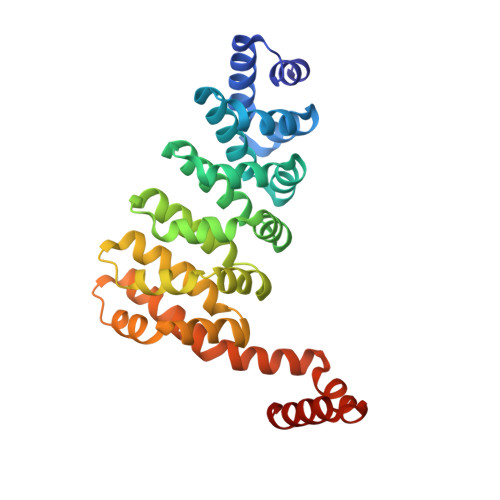
    Designed armadillo repeat proteins (dArmRP) are promising modular proteins for the engineering of binding molecules that recognize extended polypeptide chains. We determined the structure of a dArmRP containing five internal repeats and 3rd generation capping repeats in three different states by X-ray crystallography: without N-terminal His6 -tag and in the presence of calcium (YM5 A/Ca(2+) ), without N-terminal His6 -tag and in the absence of calcium (YM5 A), and with N-terminal His6 -tag and in the presence of calcium (His-YM5 A/Ca(2+)). All structures show different quaternary structures and superhelical parameters. His-YM5 A/Ca(2+) forms a crystallographic dimer, which is bridged by the His6 -tag, YM5 A/Ca(2+) forms a domain-swapped tetramer, and only in the absence of calcium and the His6 -tag, YM5 A forms a monomer. The changes of superhelical parameters are a consequence of calcium binding, because calcium ions interact with negatively charged residues, which can also participate in the modulation of helix dipole moments between adjacent repeats. These observations are important for further optimizations of dArmRPs and provide a general illustration of how construct design and crystallization conditions can influence the exact structure of the investigated protein.


  • Organizational Affiliation

    Department of Biochemistry, University of Zürich, Winterthurerstrasse 190, 8057, Zürich, Switzerland.


Macromolecules
Find similar proteins by:  (by identity cutoff)  |  3D Structure
Entity ID: 1
MoleculeChains Sequence LengthOrganismDetailsImage
Arm00010
A, B, C, D
281synthetic constructMutation(s): 0 
Entity Groups  
Sequence Clusters30% Identity50% Identity70% Identity90% Identity95% Identity100% Identity
Sequence Annotations
Expand
  • Reference Sequence
Small Molecules
Ligands 2 Unique
IDChains Name / Formula / InChI Key2D Diagram3D Interactions
ACT
Query on ACT

Download Ideal Coordinates CCD File 
N [auth B],
P [auth C]
ACETATE ION
C2 H3 O2
QTBSBXVTEAMEQO-UHFFFAOYSA-M
CA
Query on CA

Download Ideal Coordinates CCD File 
E [auth A]
F [auth A]
G [auth A]
H [auth A]
I [auth A]
E [auth A],
F [auth A],
G [auth A],
H [auth A],
I [auth A],
J [auth B],
K [auth B],
L [auth B],
M [auth B],
O [auth B]
CALCIUM ION
Ca
BHPQYMZQTOCNFJ-UHFFFAOYSA-N
Experimental Data & Validation

Experimental Data

  • Method: X-RAY DIFFRACTION
  • Resolution: 2.35 Å
  • R-Value Free: 0.267 
  • R-Value Work: 0.183 
  • R-Value Observed: 0.187 
  • Space Group: P 21 21 21
Unit Cell:
Length ( Å )Angle ( ˚ )
a = 63.87α = 90
b = 95.86β = 90
c = 177.07γ = 90
Software Package:
Software NamePurpose
REFMACrefinement
XDSdata reduction
PDB_EXTRACTdata extraction
XSCALEdata scaling
PHASERphasing

Structure Validation

View Full Validation Report


Entry History 

Deposition Data

Revision History  (Full details and data files)

  • Version 1.0: 2014-12-10
    Type: Initial release
  • Version 1.1: 2023-12-20
    Changes: Data collection, Database references, Derived calculations, Refinement description