4P5F

The crystal structure of type III effector protein XopQ complexed with adenosine diphosphate ribose


Experimental Data Snapshot

  • Method: X-RAY DIFFRACTION
  • Resolution: 2.10 Å
  • R-Value Free: 0.253 
  • R-Value Work: 0.202 
  • R-Value Observed: 0.204 

wwPDB Validation   3D Report Full Report


Ligand Structure Quality Assessment 


This is version 1.4 of the entry. See complete history


Literature

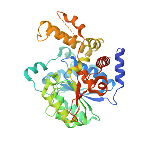
The crystal structure of type III effector protein XopQ from Xanthomonas oryzae complexed with adenosine diphosphate ribose.

Yu, S.Hwang, I.Rhee, S.

(2014) Proteins 82: 2910-2914

  • DOI: https://doi.org/10.1002/prot.24656
  • Primary Citation of Related Structures:  
    4P5F

  • PubMed Abstract: 

    Effector proteins are virulence factors that promote pathogenesis by interfering with various cellular events and are delivered directly into host cells by the secretion systems of many Gram-negative bacteria. Type III effector protein XOO4466 from the plant pathogen Xanthomonas oryzae pv. oryzae (XopQ(Xoo)) and XopQ homologs from other phytopathogens have been predicted to be nucleoside hydrolases based on their sequence similarities. However, despite such similarities, recent structural and functional studies have revealed that XopQ(Xoo) does not exhibit the expected activity of a nucleoside hydrolase. On the basis of the conservation of a Ca(2+) coordination shell of a ribose-binding site and the spacious active site in XopQ(Xoo), we hypothesized that a novel compound containing a ribosyl moiety could serve as a substrate for XopQ(Xoo). Here, we report the crystal structure of XopQ(Xoo) in complex with adenosine diphosphate ribose (ADPR), which is involved in regulating cytoplasmic Ca(2+) concentrations in eukaryotic cells. ADPR is bound to the active site of XopQ(Xoo) with its ribosyl end tethered to the Ca(2+) coordination shell. The binding of ADPR is further stabilized by interactions mediated by hydrophobic residues that undergo ligand-induced conformational changes. These data showed that XopQ(Xoo) is capable of binding a novel chemical bearing a ribosyl moiety, thereby providing the first step toward understanding the functional role of XopQ(Xoo).


  • Organizational Affiliation

    Department of Agricultural Biotechnology, Seoul National University, Seoul, Korea.


Macromolecules
Find similar proteins by:  (by identity cutoff)  |  3D Structure
Entity ID: 1
MoleculeChains Sequence LengthOrganismDetailsImage
Inosine-uridine nucleoside N-ribohydrolase
A, B
383Xanthomonas oryzae pv. oryzae KACC 10331Mutation(s): 0 
Gene Names: URH1XOO4466
UniProt
Find proteins for Q5GUA3 (Xanthomonas oryzae pv. oryzae (strain KACC10331 / KXO85))
Explore Q5GUA3 
Go to UniProtKB:  Q5GUA3
Entity Groups  
Sequence Clusters30% Identity50% Identity70% Identity90% Identity95% Identity100% Identity
UniProt GroupQ5GUA3
Sequence Annotations
Expand
  • Reference Sequence
Small Molecules
Ligands 2 Unique
IDChains Name / Formula / InChI Key2D Diagram3D Interactions
AR6
Query on AR6

Download Ideal Coordinates CCD File 
F [auth B][(2R,3S,4R,5R)-5-(6-AMINOPURIN-9-YL)-3,4-DIHYDROXY-OXOLAN-2-YL]METHYL [HYDROXY-[[(2R,3S,4R,5S)-3,4,5-TRIHYDROXYOXOLAN-2-YL]METHOXY]PHOSPHORYL] HYDROGEN PHOSPHATE
C15 H23 N5 O14 P2
SRNWOUGRCWSEMX-ZQSHOCFMSA-N
CA
Query on CA

Download Ideal Coordinates CCD File 
C [auth A],
D [auth B],
E [auth B]
CALCIUM ION
Ca
BHPQYMZQTOCNFJ-UHFFFAOYSA-N
Experimental Data & Validation

Experimental Data

  • Method: X-RAY DIFFRACTION
  • Resolution: 2.10 Å
  • R-Value Free: 0.253 
  • R-Value Work: 0.202 
  • R-Value Observed: 0.204 
  • Space Group: P 21 21 21
Unit Cell:
Length ( Å )Angle ( ˚ )
a = 47.999α = 90
b = 73.808β = 90
c = 201.943γ = 90
Software Package:
Software NamePurpose
SCALEPACKdata scaling
PDB_EXTRACTdata extraction
PHENIXmodel building
PHENIXrefinement

Structure Validation

View Full Validation Report


Ligand Structure Quality Assessment 


Entry History 

Deposition Data

Revision History  (Full details and data files)

  • Version 1.0: 2014-08-13
    Type: Initial release
  • Version 1.1: 2014-11-05
    Changes: Database references
  • Version 1.2: 2015-02-04
    Changes: Derived calculations
  • Version 1.3: 2017-09-27
    Changes: Author supporting evidence, Database references, Derived calculations, Other, Refinement description, Source and taxonomy
  • Version 1.4: 2023-12-27
    Changes: Data collection, Database references, Derived calculations, Refinement description, Structure summary