4P1Z

Structure of the MID domain from MIWI


Experimental Data Snapshot

  • Method: X-RAY DIFFRACTION
  • Resolution: 2.30 Å
  • R-Value Free: 0.275 
  • R-Value Work: 0.242 
  • R-Value Observed: 0.244 

wwPDB Validation   3D Report Full Report


This is version 1.4 of the entry. See complete history


Literature

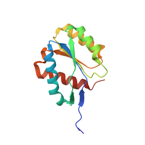
The MID-PIWI module of Piwi proteins specifies nucleotide- and strand-biases of piRNAs.

Cora, E.Pandey, R.R.Xiol, J.Taylor, J.Sachidanandam, R.McCarthy, A.A.Pillai, R.S.

(2014) RNA 20: 773-781

  • DOI: https://doi.org/10.1261/rna.044701.114
  • Primary Citation of Related Structures:  
    4P1Z

  • PubMed Abstract: 

    Piwi-interacting RNAs (piRNAs) guide Piwi Argonautes to suppress transposon activity in animal gonads. Known piRNA populations are extremely complex, with millions of individual sequences present in a single organism. Despite this complexity, specific Piwi proteins incorporate piRNAs with distinct nucleotide- and transposon strand-biases (antisense or sense) of unknown origin. Here, we examined the contribution of structural domains in Piwi proteins toward defining these biases. We report the first crystal structure of the MID domain from a Piwi Argonaute and use docking experiments to show its ability to specify recognition of 5' uridine (1U-bias) of piRNAs. Mutational analyses reveal the importance of 5' end-recognition within the MID domain for piRNA biogenesis in vivo. Finally, domain-swapping experiments uncover an unexpected role for the MID-PIWI module of a Piwi protein in dictating the transposon strand-orientation of its bound piRNAs. Our work identifies structural features that allow distinguishing individual Piwi members during piRNA biogenesis.


  • Organizational Affiliation

    European Molecular Biology Laboratory, Grenoble Outstation, 38042 Grenoble, France Unit for Virus Host-Cell Interactions, University of Grenoble Alpes-EMBL-CNRS, 38042 Grenoble, France.


Macromolecules
Find similar proteins by:  (by identity cutoff)  |  3D Structure
Entity ID: 1
MoleculeChains Sequence LengthOrganismDetailsImage
Piwi-like protein 1
A, B, C, D
128Mus musculusMutation(s): 0 
Gene Names: Piwil1Miwi
UniProt
Find proteins for Q9JMB7 (Mus musculus)
Explore Q9JMB7 
Go to UniProtKB:  Q9JMB7
Entity Groups  
Sequence Clusters30% Identity50% Identity70% Identity90% Identity95% Identity100% Identity
UniProt GroupQ9JMB7
Sequence Annotations
Expand
  • Reference Sequence
Experimental Data & Validation

Experimental Data

  • Method: X-RAY DIFFRACTION
  • Resolution: 2.30 Å
  • R-Value Free: 0.275 
  • R-Value Work: 0.242 
  • R-Value Observed: 0.244 
  • Space Group: P 21 21 21
Unit Cell:
Length ( Å )Angle ( ˚ )
a = 35.86α = 90
b = 101.29β = 90
c = 146.48γ = 90
Software Package:
Software NamePurpose
XDSdata reduction
SCALAdata scaling
SHELXDEphasing
SHARPphasing
REFMACrefinement
PDB_EXTRACTdata extraction
SHARPphasing
XSCALEdata scaling

Structure Validation

View Full Validation Report


Entry History 

Deposition Data

Revision History  (Full details and data files)

  • Version 1.0: 2014-05-07
    Type: Initial release
  • Version 1.1: 2014-10-01
    Changes: Database references
  • Version 1.2: 2015-09-23
    Changes: Data collection
  • Version 1.3: 2015-09-30
    Changes: Data collection
  • Version 1.4: 2023-12-27
    Changes: Data collection, Database references, Derived calculations, Refinement description