4P18

Crystal Structure of frog M ferritin mutant D80K


Experimental Data Snapshot

  • Method: X-RAY DIFFRACTION
  • Resolution: 1.91 Å
  • R-Value Free: 0.231 
  • R-Value Work: 0.196 
  • R-Value Observed: 0.198 

wwPDB Validation   3D Report Full Report


This is version 1.3 of the entry. See complete history


Literature

Loop electrostatics modulates the intersubunit interactions in ferritin.

Bernacchioni, C.Ghini, V.Pozzi, C.Di Pisa, F.Theil, E.C.Turano, P.

(2014) ACS Chem Biol 9: 2517-2525

  • DOI: https://doi.org/10.1021/cb500431r
  • Primary Citation of Related Structures:  
    4P18

  • PubMed Abstract: 

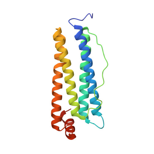
    Functional ferritins are 24-mer nanocages that self-assemble with extended contacts between pairs of 4-helix bundle subunits coupled in an antiparallel fashion along the C2 axes. The largest intersubunit interaction surface in the ferritin nanocage involves helices, but contacts also occur between groups of three residues midway in the long, solvent-exposed L-loops of facing subunits. The anchor points between intersubunit L-loop pairs are the salt bridges between the symmetry-related, conserved residues Asp80 and Lys82. The resulting quaternary structure of the cage is highly soluble and thermostable. Substitution of negatively charged Asp80 with a positively charged Lys in homopolymeric M ferritin introduces electrostatic repulsions that inhibit the oligomerization of the ferritin subunits. D80K ferritin was present in inclusion bodies under standard overexpressing conditions in E. coli, contrasting with the wild type protein. Small amounts of fully functional D80K nanocages formed when expression was slowed. The more positively charged surface results in a different solubility profile and D80K crystallized in a crystal form with a low density packing. The 3D structure of D80K variant is the same as wild type except for the side chain orientations of Lys80 and facing Lys82. When three contiguous Lys groups are introduced in D80KI81K ferritin variant the nanocage assembly is further inhibited leading to lower solubility and reduced thermal stability. Here, we demonstrate that the electrostatic pairing at the center of the L-loops has a specific kinetic role in the self-assembly of ferritin nanocages.


  • Organizational Affiliation

    Magnetic Resonance Center CERM, University of Florence , Via Luigi Sacconi 6, 50019 Sesto Fiorentino, Florence, Italy.


Macromolecules
Find similar proteins by:  (by identity cutoff)  |  3D Structure
Entity ID: 1
MoleculeChains Sequence LengthOrganismDetailsImage
Ferritin, middle subunit176Aquarana catesbeianaMutation(s): 1 
EC: 1.16.3.1
UniProt
Find proteins for P07798 (Aquarana catesbeiana)
Explore P07798 
Go to UniProtKB:  P07798
Entity Groups  
Sequence Clusters30% Identity50% Identity70% Identity90% Identity95% Identity100% Identity
UniProt GroupP07798
Sequence Annotations
Expand
  • Reference Sequence
Small Molecules
Ligands 4 Unique
IDChains Name / Formula / InChI Key2D Diagram3D Interactions
SO4
Query on SO4

Download Ideal Coordinates CCD File 
BB [auth P]
GA [auth C]
ID [auth R]
JB [auth T]
JC [auth H]
BB [auth P],
GA [auth C],
ID [auth R],
JB [auth T],
JC [auth H],
JD [auth R],
KB [auth T],
KC [auth H],
LA [auth D],
MA [auth D],
MC [auth I],
OD [auth U],
QC [auth J],
RC [auth J],
SA [auth N],
TA [auth N],
TB [auth F],
UD [auth W],
VC [auth K],
VD [auth W],
WC [auth K],
XA [auth O],
Y [auth A],
YB [auth G],
ZC [auth L]
SULFATE ION
O4 S
QAOWNCQODCNURD-UHFFFAOYSA-L
EDO
Query on EDO

Download Ideal Coordinates CCD File 
AB [auth O]
BE [auth X]
CB [auth P]
CC [auth G]
CD [auth L]
AB [auth O],
BE [auth X],
CB [auth P],
CC [auth G],
CD [auth L],
CE [auth X],
DA [auth B],
DB [auth P],
DC [auth G],
DD [auth L],
EA [auth B],
EB [auth P],
EC [auth G],
FA [auth B],
FC [auth G],
FD [auth Q],
GB [auth S],
GC [auth G],
GD [auth Q],
HB [auth S],
HC [auth G],
IA [auth C],
IB [auth S],
IC [auth G],
JA [auth C],
KA [auth C],
KD [auth R],
LD [auth R],
MD [auth R],
NB [auth T],
ND [auth R],
OC [auth I],
PA [auth D],
PC [auth I],
RA [auth M],
RB [auth E],
RD [auth U],
SB [auth E],
SD [auth V],
TC [auth J],
TD [auth V],
UA [auth N],
UC [auth J],
VA [auth N],
WA [auth N],
WB [auth F],
XB [auth F],
XC [auth K],
XD [auth W],
YC [auth K],
Z [auth A]
1,2-ETHANEDIOL
C2 H6 O2
LYCAIKOWRPUZTN-UHFFFAOYSA-N
ACT
Query on ACT

Download Ideal Coordinates CCD File 
AA [auth A],
HD [auth Q]
ACETATE ION
C2 H3 O2
QTBSBXVTEAMEQO-UHFFFAOYSA-M
CL
Query on CL

Download Ideal Coordinates CCD File 
AC [auth G]
AD [auth L]
AE [auth X]
BA [auth B]
BC [auth G]
AC [auth G],
AD [auth L],
AE [auth X],
BA [auth B],
BC [auth G],
BD [auth L],
CA [auth B],
ED [auth Q],
FB [auth S],
HA [auth C],
LB [auth T],
LC [auth H],
MB [auth T],
NA [auth D],
NC [auth I],
OA [auth D],
OB [auth E],
PB [auth E],
PD [auth U],
QA [auth M],
QB [auth E],
QD [auth U],
SC [auth J],
UB [auth F],
VB [auth F],
WD [auth W],
YA [auth O],
YD [auth X],
ZA [auth O],
ZB [auth G],
ZD [auth X]
CHLORIDE ION
Cl
VEXZGXHMUGYJMC-UHFFFAOYSA-M
Experimental Data & Validation

Experimental Data

  • Method: X-RAY DIFFRACTION
  • Resolution: 1.91 Å
  • R-Value Free: 0.231 
  • R-Value Work: 0.196 
  • R-Value Observed: 0.198 
  • Space Group: C 1 2 1
Unit Cell:
Length ( Å )Angle ( ˚ )
a = 238.93α = 90
b = 238.43β = 94.33
c = 119.69γ = 90
Software Package:
Software NamePurpose
REFMACrefinement

Structure Validation

View Full Validation Report


Entry History 

Deposition Data

Revision History  (Full details and data files)

  • Version 1.0: 2014-10-01
    Type: Initial release
  • Version 1.1: 2014-12-03
    Changes: Database references
  • Version 1.2: 2017-11-01
    Changes: Advisory, Author supporting evidence, Data collection, Derived calculations, Other, Source and taxonomy
  • Version 1.3: 2023-09-27
    Changes: Data collection, Database references, Refinement description