4OW4

Beta-trefoil designed by folding nucleus symmetric expansion ("Phifoil")


Experimental Data Snapshot

  • Method: X-RAY DIFFRACTION
  • Resolution: 2.15 Å
  • R-Value Free: 0.268 
  • R-Value Work: 0.229 
  • R-Value Observed: 0.231 

wwPDB Validation   3D Report Full Report


This is version 1.1 of the entry. See complete history


Literature

Evolution and design of protein structure by folding nucleus symmetric expansion.

Longo, L.M.Kumru, O.S.Middaugh, C.R.Blaber, M.

(2014) Structure 22: 1377-1384

  • DOI: https://doi.org/10.1016/j.str.2014.08.008
  • Primary Citation of Related Structures:  
    4OW4

  • PubMed Abstract: 

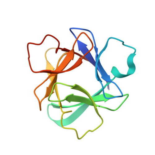
    Models of symmetric protein evolution typically invoke gene duplication and fusion events, in which repetition of a structural motif generates foldable, stable symmetric protein architecture. Success of such evolutionary processes suggests that the duplicated structural motif must be capable of nucleating protein folding. If correct, symmetric expansion of a folding nucleus sequence derived from an extant symmetric fold may be an elegant and computationally tractable solution to de novo protein design. We report the efficient de novo design of a β-trefoil protein by symmetric expansion of a β-trefoil folding nucleus, previously identified by ɸ-value analysis. The resulting protein, having exact sequence symmetry, exhibits superior folding properties compared to its naturally evolved progenitor-with the potential for redundant folding nuclei. In principle, folding nucleus symmetric expansion can be applied to any given symmetric protein fold (that is, nearly one-third of the known proteome) provided information of the folding nucleus is available.


  • Organizational Affiliation

    Department of Biomedical Sciences, Florida State University, Tallahassee, FL 32306-4300, USA.


Macromolecules
Find similar proteins by:  (by identity cutoff)  |  3D Structure
Entity ID: 1
MoleculeChains Sequence LengthOrganismDetailsImage
Beta-terfoil designed by folding nucleus symmetric expansion ("Phifoil")
A, B
123synthetic constructMutation(s): 0 
Entity Groups  
Sequence Clusters30% Identity50% Identity70% Identity90% Identity95% Identity100% Identity
Sequence Annotations
Expand
  • Reference Sequence
Small Molecules
Ligands 1 Unique
IDChains Name / Formula / InChI Key2D Diagram3D Interactions
SO4
Query on SO4

Download Ideal Coordinates CCD File 
C [auth B]SULFATE ION
O4 S
QAOWNCQODCNURD-UHFFFAOYSA-L
Experimental Data & Validation

Experimental Data

  • Method: X-RAY DIFFRACTION
  • Resolution: 2.15 Å
  • R-Value Free: 0.268 
  • R-Value Work: 0.229 
  • R-Value Observed: 0.231 
  • Space Group: P 21 21 21
Unit Cell:
Length ( Å )Angle ( ˚ )
a = 52.861α = 90
b = 68.634β = 90
c = 76.044γ = 90
Software Package:
Software NamePurpose
PDB_EXTRACTdata extraction
PHENIXrefinement

Structure Validation

View Full Validation Report


Entry History 

Deposition Data

Revision History  (Full details and data files)

  • Version 1.0: 2014-12-17
    Type: Initial release
  • Version 1.1: 2023-12-27
    Changes: Data collection, Database references, Derived calculations, Other, Refinement description, Source and taxonomy, Structure summary