4ONG

Fab fragment of 3D6 in complex with amyloid beta 1-40


Experimental Data Snapshot

  • Method: X-RAY DIFFRACTION
  • Resolution: 2.20 Å
  • R-Value Free: 0.227 
  • R-Value Work: 0.177 
  • R-Value Observed: 0.180 

wwPDB Validation   3D Report Full Report


This is version 1.3 of the entry. See complete history


Literature

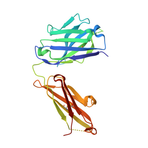
Crystal structure reveals conservation of amyloid-beta conformation recognized by 3D6 following humanization to bapineuzumab.

Feinberg, H.Saldanha, J.W.Diep, L.Goel, A.Widom, A.Veldman, G.M.Weis, W.I.Schenk, D.Basi, G.S.

(2014) Alzheimers Res Ther 6: 31-31

  • DOI: https://doi.org/10.1186/alzrt261
  • Primary Citation of Related Structures:  
    4ONF, 4ONG

  • PubMed Abstract: 

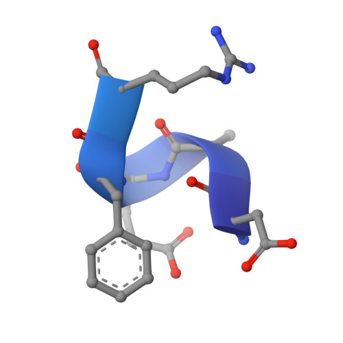
    Immunotherapy targeting amyloid-β peptide is under active clinical investigation for treatment of Alzheimer's disease (AD). Among the hypotheses being investigated for impact on clinical outcome are the preferred epitope or conformation of amyloid-β to target for treatment, and the mechanism of action underlying immunotherapy. Bapineuzumab (humanized 3D6), a neo-epitope specific antibody recognizing amyloid-β1-5 with strong preference for an exposed Asp residue at the N-terminus of the peptide, has undergone advanced clinical testing for treatment of AD. To gain further insight into the epitope conformation, we interrogated structural details of amino-terminal epitopes in amyloid-β using x-ray crystallography of 3D6Fab:amyloid-β complexes. Humanization of 3D6 was carried out using standard procedures integrating recombinant methods, sequence informatics, and homology modeling predictions to identify important mouse framework residues for retention in the finished humanized product. Here we report the crystal structure of a recombinant Fab fragment of 3D6 in complex with amyloid-β1-7 solved at 2.0 Å resolution. The N-terminus of amyloid-β is bound to 3D6 as a 310 helix. The amino-terminal Asp residue is buried deepest in the antibody binding pocket, with the Cβ atom of residue 6 visible at the entrance to the binding pocket near the surface of the antibody. We further evaluate homology model based predictions used to guide humanization of 3D6 to bapineuzumab, with actual structure of the Fab. The structure of the Fab:amyloid-β complex validates design of the humanized antibody, and confirms the amyloid-β epitope recognized by 3D6 as previously mapped by ELISA. The conformation of amyloid-β antigen recognized by 3D6 is novel and distinct from other antibodies recognizing N-terminal epitopes. Our result provides the first report demonstrating structural conservation of antigen contact residues, and conformation of antigen recognized, between the parent murine antibody and its humanized version.


  • Organizational Affiliation

    Departments of Structural Biology and of Molecular & Cellular Physiology, 299 Campus Drive, Stanford University School of Medicine, Stanford, CA 94305, USA.


Macromolecules
Find similar proteins by:  (by identity cutoff)  |  3D Structure
Entity ID: 1
MoleculeChains Sequence LengthOrganismDetailsImage
3D6 FAB ANTIBODY HEAVY CHAINA [auth H]222Mus musculusMutation(s): 0 
Entity Groups  
Sequence Clusters30% Identity50% Identity70% Identity90% Identity95% Identity100% Identity
Sequence Annotations
Expand
  • Reference Sequence
Find similar proteins by:  (by identity cutoff)  |  3D Structure
Entity ID: 2
MoleculeChains Sequence LengthOrganismDetailsImage
3D6 FAB ANTIBODY LIGHT CHAINB [auth L]219Mus musculusMutation(s): 0 
Entity Groups  
Sequence Clusters30% Identity50% Identity70% Identity90% Identity95% Identity100% Identity
Sequence Annotations
Expand
  • Reference Sequence
Find similar proteins by:  (by identity cutoff)  |  3D Structure
Entity ID: 3
MoleculeChains Sequence LengthOrganismDetailsImage
Amyloid beta A4 proteinC [auth P]40Homo sapiensMutation(s): 0 
UniProt & NIH Common Fund Data Resources
Find proteins for P05067 (Homo sapiens)
Explore P05067 
Go to UniProtKB:  P05067
PHAROS:  P05067
GTEx:  ENSG00000142192 
Entity Groups  
Sequence Clusters30% Identity50% Identity70% Identity90% Identity95% Identity100% Identity
UniProt GroupP05067
Sequence Annotations
Expand
  • Reference Sequence
Small Molecules
Ligands 3 Unique
IDChains Name / Formula / InChI Key2D Diagram3D Interactions
IMD
Query on IMD

Download Ideal Coordinates CCD File 
LA [auth L],
Q [auth H],
R [auth H]
IMIDAZOLE
C3 H5 N2
RAXXELZNTBOGNW-UHFFFAOYSA-O
ZN
Query on ZN

Download Ideal Coordinates CCD File 
AA [auth L]
BA [auth L]
CA [auth L]
D [auth H]
DA [auth L]
AA [auth L],
BA [auth L],
CA [auth L],
D [auth H],
DA [auth L],
E [auth H],
EA [auth L],
F [auth H],
FA [auth L],
G [auth H],
GA [auth L],
H,
HA [auth L],
I [auth H],
IA [auth L],
J [auth H],
JA [auth L],
K [auth H],
KA [auth L],
L [auth H],
M [auth H],
N [auth H],
O [auth H],
P [auth H],
X [auth L],
Y [auth L],
Z [auth L]
ZINC ION
Zn
PTFCDOFLOPIGGS-UHFFFAOYSA-N
CL
Query on CL

Download Ideal Coordinates CCD File 
MA [auth L]
NA [auth L]
OA [auth L]
PA [auth L]
QA [auth L]
MA [auth L],
NA [auth L],
OA [auth L],
PA [auth L],
QA [auth L],
RA [auth L],
S [auth H],
T [auth H],
U [auth H],
V [auth H],
W [auth H]
CHLORIDE ION
Cl
VEXZGXHMUGYJMC-UHFFFAOYSA-M
Experimental Data & Validation

Experimental Data

  • Method: X-RAY DIFFRACTION
  • Resolution: 2.20 Å
  • R-Value Free: 0.227 
  • R-Value Work: 0.177 
  • R-Value Observed: 0.180 
  • Space Group: P 2 2 21
Unit Cell:
Length ( Å )Angle ( ˚ )
a = 40.04α = 90
b = 84.94β = 90
c = 175.69γ = 90
Software Package:
Software NamePurpose
MOSFLMdata reduction
SCALAdata scaling
PHENIXrefinement
PDB_EXTRACTdata extraction
Blu-Icedata collection
COMOphasing

Structure Validation

View Full Validation Report


Entry History 

Deposition Data

Revision History  (Full details and data files)

  • Version 1.0: 2014-06-11
    Type: Initial release
  • Version 1.1: 2014-07-30
    Changes: Database references
  • Version 1.2: 2017-11-22
    Changes: Refinement description
  • Version 1.3: 2024-04-03
    Changes: Data collection, Database references, Derived calculations, Refinement description