4OIT

Structure, interactions and evolutionary implications of a domain-swapped lectin dimer from Mycobacterium smegmatis


Experimental Data Snapshot

  • Method: X-RAY DIFFRACTION
  • Resolution: 2.24 Å
  • R-Value Free: 0.261 
  • R-Value Work: 0.220 
  • R-Value Observed: 0.222 

wwPDB Validation   3D Report Full Report


Ligand Structure Quality Assessment 


This is version 1.3 of the entry. See complete history


Literature

Structure, interactions and evolutionary implications of a domain-swapped lectin dimer from Mycobacterium smegmatis.

Patra, D.Mishra, P.Surolia, A.Vijayan, M.

(2014) Glycobiology 24: 956-965

  • DOI: https://doi.org/10.1093/glycob/cwu059
  • Primary Citation of Related Structures:  
    4OIT, 4OIZ, 4OKC

  • PubMed Abstract: 

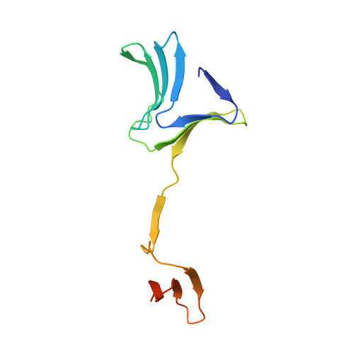
    Crystal structure determination of the lectin domain of MSMEG_3662 from Mycobacterium smegmatis and its complexes with mannose and methyl-α-mannose, the first effort of its kind on a mycobacterial lectin, reveals a structure very similar to β-prism II fold lectins from plant sources, but with extensive unprecedented domain swapping in dimer formation. The two subunits in a dimer often show small differences in structure, but the two domains, not always related by 2-fold symmetry, have the same structure. Each domain carries three sugar-binding sites, similar to those in plant lectins, one on each Greek key motif. The occurrence of β-prism II fold lectins in bacteria, with characteristics similar to those from plants, indicates that this family of lectins is of ancient origin and had evolved into a mature system before bacteria and plants diverged. In plants, the number of binding sites per domain varies between one and three, whereas the number is two in the recently reported lectin domains from Pseudomonas putida and Pseudomonas aeruginosa. An analysis of the sequences of the lectins and the lectin domains shows that the level of sequence similarity among the three Greek keys in each domain has a correlation with the number of binding sites in it. Furthermore, sequence conservation among the lectins from different species is the highest for that Greek key which carries a binding site in all of them. Thus, it would appear that carbohydrate binding influences the course of the evolution of the lectin.


  • Organizational Affiliation

    Molecular Biophysics Unit, Indian Institute of Science, Bangalore 560012, India.


Macromolecules
Find similar proteins by:  (by identity cutoff)  |  3D Structure
Entity ID: 1
MoleculeChains Sequence LengthOrganismDetailsImage
LysM domain protein
A, B, C, D
113Mycolicibacterium smegmatis MC2 155Mutation(s): 0 
Gene Names: MSMEG_3662
UniProt
Find proteins for A0QYH7 (Mycolicibacterium smegmatis (strain ATCC 700084 / mc(2)155))
Explore A0QYH7 
Go to UniProtKB:  A0QYH7
Entity Groups  
Sequence Clusters30% Identity50% Identity70% Identity90% Identity95% Identity100% Identity
UniProt GroupA0QYH7
Sequence Annotations
Expand
  • Reference Sequence
Small Molecules
Ligands 2 Unique
IDChains Name / Formula / InChI Key2D Diagram3D Interactions
BMA
Query on BMA

Download Ideal Coordinates CCD File 
I [auth B]beta-D-mannopyranose
C6 H12 O6
WQZGKKKJIJFFOK-RWOPYEJCSA-N
MAN
Query on MAN

Download Ideal Coordinates CCD File 
E [auth A]
F [auth A]
G [auth A]
H [auth B]
J [auth C]
E [auth A],
F [auth A],
G [auth A],
H [auth B],
J [auth C],
K [auth C],
L [auth C],
M [auth D],
N [auth D],
O [auth D]
alpha-D-mannopyranose
C6 H12 O6
WQZGKKKJIJFFOK-PQMKYFCFSA-N
Binding Affinity Annotations 
IDSourceBinding Affinity
MAN Binding MOAD:  4OIT Ka: 32.5 (M^-1) from 1 assay(s)
Experimental Data & Validation

Experimental Data

  • Method: X-RAY DIFFRACTION
  • Resolution: 2.24 Å
  • R-Value Free: 0.261 
  • R-Value Work: 0.220 
  • R-Value Observed: 0.222 
  • Space Group: P 1 21 1
Unit Cell:
Length ( Å )Angle ( ˚ )
a = 47.03α = 90
b = 80.08β = 105.98
c = 56.91γ = 90
Software Package:
Software NamePurpose
MAR345dtbdata collection
PHASERphasing
REFMACrefinement
MOSFLMdata reduction
SCALAdata scaling

Structure Validation

View Full Validation Report


Ligand Structure Quality Assessment 


Entry History 

Deposition Data

Revision History  (Full details and data files)

  • Version 1.0: 2014-07-23
    Type: Initial release
  • Version 1.1: 2020-07-29
    Type: Remediation
    Reason: Carbohydrate remediation
    Changes: Data collection, Database references, Derived calculations, Structure summary
  • Version 1.2: 2022-08-24
    Changes: Database references, Structure summary
  • Version 1.3: 2024-04-03
    Changes: Data collection, Refinement description