4OIF

3D structure of Gan42B, a GH42 beta-galactosidase from G.


Experimental Data Snapshot

  • Method: X-RAY DIFFRACTION
  • Resolution: 2.45 Å
  • R-Value Free: 0.204 
  • R-Value Work: 0.133 
  • R-Value Observed: 0.137 

wwPDB Validation   3D Report Full Report


This is version 1.3 of the entry. See complete history


Literature

Crystallization and preliminary crystallographic analysis of GanB, a GH42 intracellular beta-galactosidase from Geobacillus stearothermophilus.

Solomon, H.V.Tabachnikov, O.Feinberg, H.Govada, L.Chayen, N.E.Shoham, Y.Shoham, G.

(2013) Acta Crystallogr Sect F Struct Biol Cryst Commun 69: 1114-1119

  • DOI: https://doi.org/10.1107/S1744309113023609
  • Primary Citation of Related Structures:  
    4OIF

  • PubMed Abstract: 

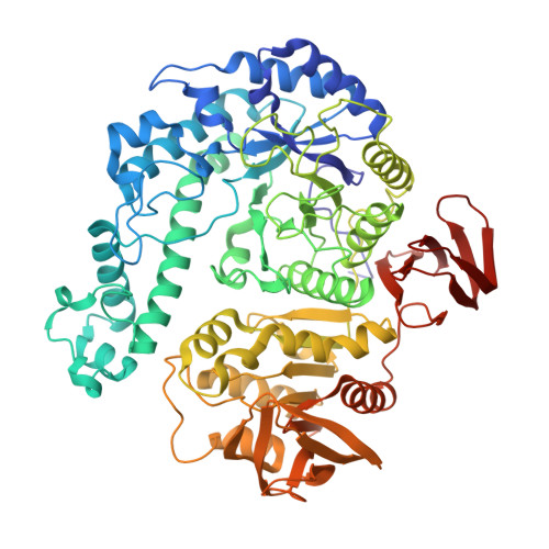
    Geobacillus stearothermophilus T-6 is a Gram-positive thermophilic soil bacterium that contains a multi-enzyme system for the utilization of plant cell-wall polysaccharides, including xylan, arabinan and galactan. The bacterium uses a number of endo-acting extracellular enzymes that break down the high-molecular-weight polysaccharides into decorated oligosaccharides. These oligosaccharides enter the cell and are further hydrolyzed into sugar monomers by a set of intracellular glycoside hydrolases. One of these intracellular degrading enzymes is GanB, a glycoside hydrolase family 42 β-galactosidase capable of hydrolyzing short β-1,4-galactosaccharides to galactose. GanB and related enzymes therefore play an important part in the hemicellulolytic utilization system of many microorganisms which use plant biomass for growth. The interest in the biochemical characterization and structural analysis of these enzymes stems from their potential biotechnological applications. GanB from G. stearothermophilus T-6 has recently been cloned, overexpressed, purified, biochemically characterized and crystallized in our laboratory as part of its complete structure-function study. The best crystals obtained for this enzyme belong to the primitive orthorhombic space group P2₁2₁2₁, with average crystallographic unit-cell parameters of a=71.84, b=181.35, c=196.57 Å. Full diffraction data sets to 2.45 and 2.50 Å resolution have been collected for both the wild-type enzyme and its E323A nucleophile catalytic mutant, respectively, as measured from flash-cooled crystals at 100 K using synchrotron radiation. These data are currently being used for the full three-dimensional crystal structure determination of GanB.


  • Organizational Affiliation

    Institute of Chemistry and the Laboratory for Structural Chemistry and Biology, The Hebrew University of Jerusalem, Jerusalem 91904, Israel.


Macromolecules
Find similar proteins by:  (by identity cutoff)  |  3D Structure
Entity ID: 1
MoleculeChains Sequence LengthOrganismDetailsImage
Beta-galactosidase
A, B, C
686Geobacillus stearothermophilusMutation(s): 0 
Gene Names: gan42BganB
EC: 3.2.1.23
UniProt
Find proteins for F8TRX0 (Geobacillus stearothermophilus)
Explore F8TRX0 
Go to UniProtKB:  F8TRX0
Entity Groups  
Sequence Clusters30% Identity50% Identity70% Identity90% Identity95% Identity100% Identity
UniProt GroupF8TRX0
Sequence Annotations
Expand
  • Reference Sequence
Small Molecules
Ligands 2 Unique
IDChains Name / Formula / InChI Key2D Diagram3D Interactions
GOL
Query on GOL

Download Ideal Coordinates CCD File 
AA [auth C]
BA [auth C]
CA [auth C]
DA [auth C]
E [auth A]
AA [auth C],
BA [auth C],
CA [auth C],
DA [auth C],
E [auth A],
EA [auth C],
F [auth A],
G [auth A],
H [auth A],
I [auth A],
J [auth A],
K [auth A],
L [auth A],
N [auth B],
O [auth B],
P [auth B],
Q [auth B],
R [auth B],
S [auth B],
T [auth B],
U [auth B],
V [auth B],
W [auth B],
X [auth B],
Y [auth B]
GLYCEROL
C3 H8 O3
PEDCQBHIVMGVHV-UHFFFAOYSA-N
ZN
Query on ZN

Download Ideal Coordinates CCD File 
D [auth A],
M [auth B],
Z [auth C]
ZINC ION
Zn
PTFCDOFLOPIGGS-UHFFFAOYSA-N
Experimental Data & Validation

Experimental Data

  • Method: X-RAY DIFFRACTION
  • Resolution: 2.45 Å
  • R-Value Free: 0.204 
  • R-Value Work: 0.133 
  • R-Value Observed: 0.137 
  • Space Group: P 21 21 21
Unit Cell:
Length ( Å )Angle ( ˚ )
a = 71.84α = 90
b = 181.35β = 90
c = 196.57γ = 90
Software Package:
Software NamePurpose
PHENIXrefinement
REFMACrefinement
ADSCdata collection
MOSFLMdata reduction
SCALAdata scaling
REFMACphasing

Structure Validation

View Full Validation Report


Entry History 

Deposition Data

Revision History  (Full details and data files)

  • Version 1.0: 2015-02-04
    Type: Initial release
  • Version 1.1: 2017-11-22
    Changes: Refinement description
  • Version 1.2: 2019-07-17
    Changes: Data collection, Refinement description
  • Version 1.3: 2023-09-20
    Changes: Data collection, Database references, Derived calculations, Refinement description