4OHB

Crystal structure of MilB E103A in complex with 5-hydroxymethylcytidine 5'-monophosphate (hmCMP) from Streptomyces rimofaciens


Experimental Data Snapshot

  • Method: X-RAY DIFFRACTION
  • Resolution: 2.40 Å
  • R-Value Free: 0.257 
  • R-Value Work: 0.192 
  • R-Value Observed: 0.195 

wwPDB Validation   3D Report Full Report


Ligand Structure Quality Assessment 


This is version 1.3 of the entry. See complete history


Literature

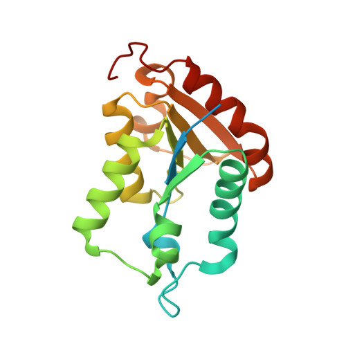
Structure of the N-glycosidase MilB in complex with hydroxymethyl CMP reveals its Arg23 specifically recognizes the substrate and controls its entry

Zhao, G.Wu, G.Zhang, Y.Liu, G.Han, T.Deng, Z.He, X.

(2014) Nucleic Acids Res 42: 8115-8124

  • DOI: https://doi.org/10.1093/nar/gku486
  • Primary Citation of Related Structures:  
    4OHB, 4OHR

  • PubMed Abstract: 

    5-Hydroxymethylcytosine (5hmC) is present in T-even phage and mammalian DNA as well as some nucleoside antibiotics, including mildiomycin and bacimethrin, during whose synthesis 5hmC is produced by the hydrolysis of 5-hydroxymethyl cytidine 5'-monophosphate (hmCMP) by an N-glycosidase MilB. Recently, the MilB-CMP complex structure revealed its substrate specificity for CMP over dCMP. However, hmCMP instead of CMP is the preferred substrate for MilB as supported by that its KM for CMP is ∼27-fold higher than that for hmCMP. Here, we determined the crystal structures of MilB and its catalytically inactive E103A mutant in complex with hmCMP. In the structure of the complex, Phe22 and Arg23 are positioned in a cage-like active site resembling the binding pocket for the flipped 5-methylcytosine (5mC) in eukaryotic 5mC-binding proteins. Van der Waals interaction between the benzene ring of Phe22 and the pyrimidine ring of hmCMP stabilizes its binding. Remarkably, upon hmCMP binding, the guanidinium group of Arg23 was bent ∼65° toward hmCMP to recognize its 5-hydroxymethyl group, inducing semi-closure of the cage-like pocket. Mutagenesis studies of Arg23 and bioinformatics analysis demonstrate that the positively charged Arg/Lys at this site is critical for the specific recognition of the 5-hydroxymethyl group of hmCMP.


  • Organizational Affiliation

    State Key Laboratory of Microbial Metabolism and School of Life Sciences and Biotechnology, Shanghai Jiao Tong University, 1954 Huashan Road, Shanghai 200030, China.


Macromolecules
Find similar proteins by:  (by identity cutoff)  |  3D Structure
Entity ID: 1
MoleculeChains Sequence LengthOrganismDetailsImage
CMP/hydroxymethyl CMP hydrolase190Streptomyces rimofaciensMutation(s): 1 
Gene Names: MilB
UniProt
Find proteins for B4Y381 (Streptomyces rimofaciens)
Explore B4Y381 
Go to UniProtKB:  B4Y381
Entity Groups  
Sequence Clusters30% Identity50% Identity70% Identity90% Identity95% Identity100% Identity
UniProt GroupB4Y381
Sequence Annotations
Expand
  • Reference Sequence
Small Molecules
Ligands 1 Unique
IDChains Name / Formula / InChI Key2D Diagram3D Interactions
5HM
Query on 5HM

Download Ideal Coordinates CCD File 
B [auth A]5-(hydroxymethyl)cytidine 5'-(dihydrogen phosphate)
C10 H16 N3 O9 P
KPUOHXMVCZBWQC-JXOAFFINSA-N
Experimental Data & Validation

Experimental Data

  • Method: X-RAY DIFFRACTION
  • Resolution: 2.40 Å
  • R-Value Free: 0.257 
  • R-Value Work: 0.192 
  • R-Value Observed: 0.195 
  • Space Group: C 2 2 21
Unit Cell:
Length ( Å )Angle ( ˚ )
a = 45.426α = 90
b = 101.808β = 90
c = 81.744γ = 90
Software Package:
Software NamePurpose
DENZOdata reduction
SCALEPACKdata scaling
REFMACrefinement
PDB_EXTRACTdata extraction

Structure Validation

View Full Validation Report


Ligand Structure Quality Assessment 


Entry History 

Deposition Data

Revision History  (Full details and data files)

  • Version 1.0: 2014-06-25
    Type: Initial release
  • Version 1.1: 2016-10-19
    Changes: Database references, Structure summary
  • Version 1.2: 2017-11-22
    Changes: Refinement description
  • Version 1.3: 2024-03-20
    Changes: Data collection, Database references, Derived calculations