4NYT

L-Ficolin Complexed to Phosphocholine


Experimental Data Snapshot

  • Method: X-RAY DIFFRACTION
  • Resolution: 2.25 Å
  • R-Value Free: 0.243 
  • R-Value Work: 0.213 
  • R-Value Observed: 0.214 

wwPDB Validation   3D Report Full Report


Ligand Structure Quality Assessment 


This is version 2.0 of the entry. See complete history


Literature

Human L-ficolin recognizes phosphocholine moieties of pneumococcal teichoic Acid

Vassal-Stermann, E.Lacroix, M.Gout, E.Laffly, E.Pedersen, C.M.Martin, L.Amoroso, A.Schmidt, R.R.Zahringer, U.Gaboriaud, C.Di Guilmi, A.M.Thielens, N.M.

(2014) J Immunol 193: 5699-5708

  • DOI: https://doi.org/10.4049/jimmunol.1400127
  • Primary Citation of Related Structures:  
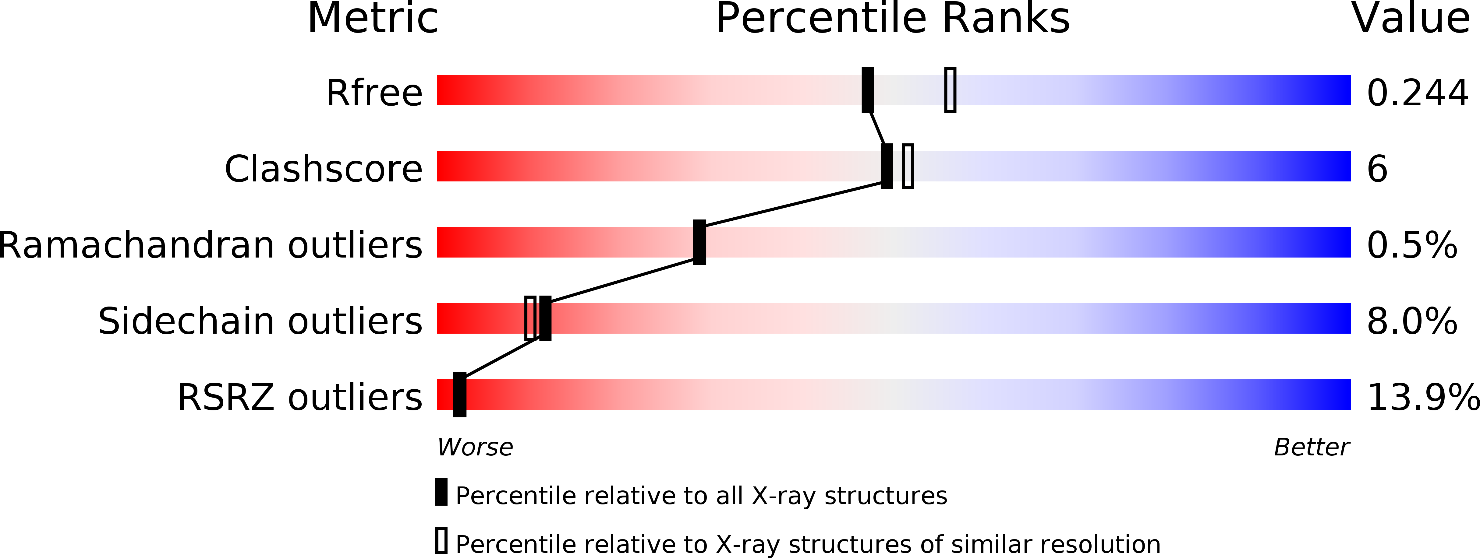
    4NYT

  • PubMed Abstract: 

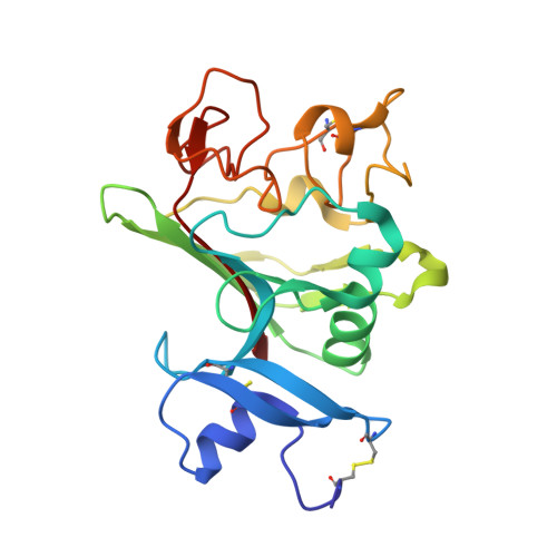
    Human L-ficolin is a soluble protein of the innate immune system able to sense pathogens through its fibrinogen (FBG) recognition domains and to trigger activation of the lectin complement pathway through associated serine proteases. L-Ficolin has been previously shown to recognize pneumococcal clinical isolates, but its ligands and especially its molecular specificity remain to be identified. Using solid-phase binding assays, serum and recombinant L-ficolins were shown to interact with serotype 2 pneumococcal strain D39 and its unencapsulated R6 derivative. Incubation of both strains with serum triggered complement activation, as measured by C4b and C3b deposition, which was decreased by using ficolin-depleted serum. Recombinant L-ficolin and its FBG-like recognition domain bound to isolated pneumococcal cell wall extracts, whereas binding to cell walls depleted of teichoic acid (TA) was decreased. Both proteins were also shown to interact with two synthetic TA compounds, each comprising part structures of the complete lipoteichoic acid molecule with two PCho residues. Competition studies and direct interaction measurements by surface plasmon resonance identified PCho as a novel L-ficolin ligand. Structural analysis of complexes of the FBG domain of L-ficolin and PCho revealed that the phosphate moiety interacts with amino acids previously shown to define an acetyl binding site. Consequently, binding of L-ficolin to immobilized acetylated BSA was inhibited by PCho and synthetic TA. Binding of serum L-ficolin to immobilized synthetic TA and PCho-conjugated BSA triggered activation of the lectin complement pathway, thus further supporting the hypothesis of L-ficolin involvement in host antipneumococcal defense.


  • Organizational Affiliation

    University of Grenoble Alpes, Institut de Biologie Structurale, F-38044 Grenoble, France; Centre National de la Recherche Scientifique, Institut de Biologie Structurale, F-38044 Grenoble, France; Commissariat à l'Energie Atomique et aux Energies Alternatives, Institut de Biologie Structurale, F-38044 Grenoble, France;


Macromolecules
Find similar proteins by:  (by identity cutoff)  |  3D Structure
Entity ID: 1
MoleculeChains Sequence LengthOrganismDetailsImage
Ficolin-2A [auth C],
B [auth A],
C [auth B]
217Homo sapiensMutation(s): 0 
Gene Names: FCN2
UniProt & NIH Common Fund Data Resources
Find proteins for Q15485 (Homo sapiens)
Explore Q15485 
Go to UniProtKB:  Q15485
PHAROS:  Q15485
GTEx:  ENSG00000160339 
Entity Groups  
Sequence Clusters30% Identity50% Identity70% Identity90% Identity95% Identity100% Identity
UniProt GroupQ15485
Sequence Annotations
Expand
  • Reference Sequence
Oligosaccharides

Help

Entity ID: 2
MoleculeChains Length2D Diagram Glycosylation3D Interactions
alpha-D-mannopyranose-(1-4)-2-acetamido-2-deoxy-beta-D-glucopyranose-(1-4)-[alpha-L-fucopyranose-(1-6)]2-acetamido-2-deoxy-beta-D-glucopyranose
D
4N-Glycosylation
Glycosylation Resources
GlyTouCan:  G47477HI
GlyCosmos:  G47477HI
GlyGen:  G47477HI
Small Molecules
Ligands 5 Unique
IDChains Name / Formula / InChI Key2D Diagram3D Interactions
NAG
Query on NAG

Download Ideal Coordinates CCD File 
I [auth A]2-acetamido-2-deoxy-beta-D-glucopyranose
C8 H15 N O6
OVRNDRQMDRJTHS-FMDGEEDCSA-N
PC
Query on PC

Download Ideal Coordinates CCD File 
J [auth A]PHOSPHOCHOLINE
C5 H15 N O4 P
YHHSONZFOIEMCP-UHFFFAOYSA-O
PO4
Query on PO4

Download Ideal Coordinates CCD File 
G [auth C]PHOSPHATE ION
O4 P
NBIIXXVUZAFLBC-UHFFFAOYSA-K
ACT
Query on ACT

Download Ideal Coordinates CCD File 
E [auth C],
H [auth A]
ACETATE ION
C2 H3 O2
QTBSBXVTEAMEQO-UHFFFAOYSA-M
CA
Query on CA

Download Ideal Coordinates CCD File 
F [auth C],
K [auth A],
L [auth A],
M [auth B]
CALCIUM ION
Ca
BHPQYMZQTOCNFJ-UHFFFAOYSA-N
Binding Affinity Annotations 
IDSourceBinding Affinity
PC Binding MOAD:  4NYT Kd: 3.70e+6 (nM) from 1 assay(s)
Experimental Data & Validation

Experimental Data

  • Method: X-RAY DIFFRACTION
  • Resolution: 2.25 Å
  • R-Value Free: 0.243 
  • R-Value Work: 0.213 
  • R-Value Observed: 0.214 
  • Space Group: P 32 2 1
Unit Cell:
Length ( Å )Angle ( ˚ )
a = 96.13α = 90
b = 96.13β = 90
c = 139.88γ = 120
Software Package:
Software NamePurpose
ADSCdata collection
PHASERphasing
REFMACrefinement
XDSdata reduction
XSCALEdata scaling

Structure Validation

View Full Validation Report


Ligand Structure Quality Assessment 


Entry History 

Deposition Data

Revision History  (Full details and data files)

  • Version 1.0: 2014-10-29
    Type: Initial release
  • Version 1.1: 2014-12-24
    Changes: Database references
  • Version 2.0: 2020-07-29
    Type: Remediation
    Reason: Carbohydrate remediation
    Changes: Advisory, Atomic model, Data collection, Derived calculations, Structure summary