4NEV

Crystal structure of Trypanothione Reductase from Trypanosoma brucei in complex with inhibitor EP127 (5-{5-[1-(PYRROLIDIN-1-YL)CYCLOHEXYL]-1,3-THIAZOL-2-YL}-1H-INDOLE)


Experimental Data Snapshot

  • Method: X-RAY DIFFRACTION
  • Resolution: 2.50 Å
  • R-Value Free: 0.258 
  • R-Value Work: 0.234 
  • R-Value Observed: 0.235 

wwPDB Validation   3D Report Full Report


Ligand Structure Quality Assessment 


This is version 1.2 of the entry. See complete history


Literature

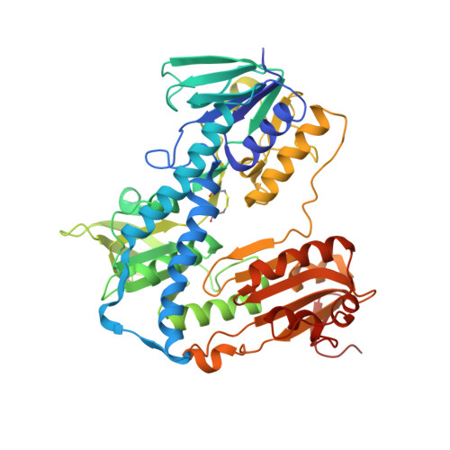
Binding to large enzyme pockets: small-molecule inhibitors of trypanothione reductase.

Persch, E.Bryson, S.Todoroff, N.K.Eberle, C.Thelemann, J.Dirdjaja, N.Kaiser, M.Weber, M.Derbani, H.Brun, R.Schneider, G.Pai, E.F.Krauth-Siegel, R.L.Diederich, F.

(2014) ChemMedChem 9: 1880-1891

  • DOI: https://doi.org/10.1002/cmdc.201402032
  • Primary Citation of Related Structures:  
    4NEV, 4NEW

  • PubMed Abstract: 

    The causative agents of the parasitic disease human African trypanosomiasis belong to the family of trypanosomatids. These parasitic protozoa exhibit a unique thiol redox metabolism that is based on the flavoenzyme trypanothione reductase (TR). TR was identified as a potential drug target and features a large active site that allows a multitude of possible ligand orientations, which renders rational structure-based inhibitor design highly challenging. Herein we describe the synthesis, binding properties, and kinetic analysis of a new series of small-molecule inhibitors of TR. The conjunction of biological activities, mutation studies, and virtual ligand docking simulations led to the prediction of a binding mode that was confirmed by crystal structure analysis. The crystal structures revealed that the ligands bind to the hydrophobic wall of the so-called "mepacrine binding site". The binding conformation and potency of the inhibitors varied for TR from Trypanosoma brucei and T. cruzi.


  • Organizational Affiliation

    Laboratorium für Organische Chemie, ETH Zürich, Vladimir-Prelog-Weg 3, 8093 Zurich (Switzerland).


Macromolecules
Find similar proteins by:  (by identity cutoff)  |  3D Structure
Entity ID: 1
MoleculeChains Sequence LengthOrganismDetailsImage
Trypanothione reductase
A, B
495Trypanosoma brucei brucei TREU927Mutation(s): 0 
Gene Names: Tb10.406.0520
EC: 1.8.1.12
UniProt
Find proteins for Q389T8 (Trypanosoma brucei brucei (strain 927/4 GUTat10.1))
Explore Q389T8 
Go to UniProtKB:  Q389T8
Entity Groups  
Sequence Clusters30% Identity50% Identity70% Identity90% Identity95% Identity100% Identity
UniProt GroupQ389T8
Sequence Annotations
Expand
  • Reference Sequence
Small Molecules
Ligands 4 Unique
IDChains Name / Formula / InChI Key2D Diagram3D Interactions
FAD
Query on FAD

Download Ideal Coordinates CCD File 
C [auth A],
H [auth B]
FLAVIN-ADENINE DINUCLEOTIDE
C27 H33 N9 O15 P2
VWWQXMAJTJZDQX-UYBVJOGSSA-N
2JR
Query on 2JR

Download Ideal Coordinates CCD File 
D [auth A],
I [auth B],
P [auth B]
5-{5-[1-(pyrrolidin-1-yl)cyclohexyl]-1,3-thiazol-2-yl}-1H-indole
C21 H25 N3 S
MXBHIYKTGVOQMD-UHFFFAOYSA-N
EPE
Query on EPE

Download Ideal Coordinates CCD File 
O [auth B]4-(2-HYDROXYETHYL)-1-PIPERAZINE ETHANESULFONIC ACID
C8 H18 N2 O4 S
JKMHFZQWWAIEOD-UHFFFAOYSA-N
SO4
Query on SO4

Download Ideal Coordinates CCD File 
E [auth A]
F [auth A]
G [auth A]
J [auth B]
K [auth B]
E [auth A],
F [auth A],
G [auth A],
J [auth B],
K [auth B],
L [auth B],
M [auth B],
N [auth B]
SULFATE ION
O4 S
QAOWNCQODCNURD-UHFFFAOYSA-L
Binding Affinity Annotations 
IDSourceBinding Affinity
2JR Binding MOAD:  4NEV Ki: 1.20e+4 (nM) from 1 assay(s)
Experimental Data & Validation

Experimental Data

  • Method: X-RAY DIFFRACTION
  • Resolution: 2.50 Å
  • R-Value Free: 0.258 
  • R-Value Work: 0.234 
  • R-Value Observed: 0.235 
  • Space Group: P 43 21 2
Unit Cell:
Length ( Å )Angle ( ˚ )
a = 117.2α = 90
b = 117.2β = 90
c = 224.6γ = 90
Software Package:
Software NamePurpose
XDSdata scaling
PHASERphasing
REFMACrefinement
HKL-2000data reduction
HKL-2000data scaling

Structure Validation

View Full Validation Report


Ligand Structure Quality Assessment 


Entry History 

Deposition Data

Revision History  (Full details and data files)

  • Version 1.0: 2014-05-14
    Type: Initial release
  • Version 1.1: 2014-08-13
    Changes: Database references
  • Version 1.2: 2023-09-20
    Changes: Data collection, Database references, Derived calculations, Refinement description