Structure of the extended-spectrum class C beta-lactamase ADC-1 from Acinetobacter baumannii.
Bhattacharya, M., Toth, M., Antunes, N.T., Smith, C.A., Vakulenko, S.B.(2014) Acta Crystallogr D Biol Crystallogr 70: 760-771
- PubMed: 24598745
- DOI: https://doi.org/10.1107/S1399004713033014
- Primary Citation of Related Structures:
4NET - PubMed Abstract:
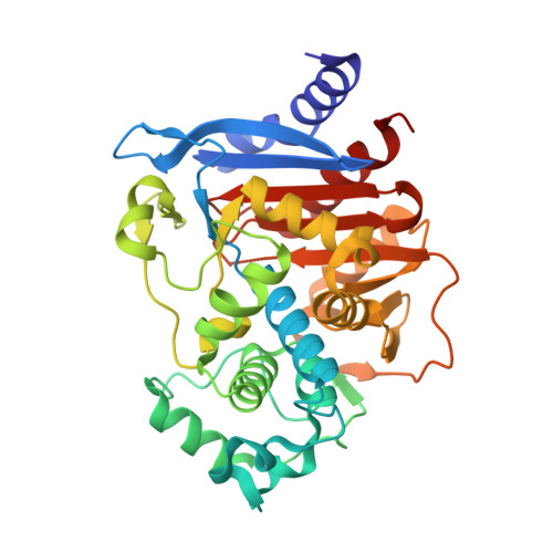
ADC-type class C β-lactamases comprise a large group of enzymes that are encoded by genes located on the chromosome of Acinetobacter baumannii, a causative agent of serious bacterial infections. Overexpression of these enzymes renders A. baumannii resistant to various β-lactam antibiotics and thus severely compromises the ability to treat infections caused by this deadly pathogen. Here, the high-resolution crystal structure of ADC-1, the first member of this clinically important family of antibiotic-resistant enzymes, is reported. Unlike the narrow-spectrum class C β-lactamases, ADC-1 is capable of producing resistance to the expanded-spectrum cephalosporins, rendering them inactive against A. baumannii. The extension of the substrate profile of the enzyme is likely to be the result of structural differences in the R2-loop, primarily the deletion of three residues and subsequent rearrangement of the A10a and A10b helices. These structural rearrangements result in the enlargement of the R2 pocket of ADC-1, allowing it to accommodate the bulky R2 substituents of the third-generation cephalosporins, thus enhancing the catalytic efficiency of the enzyme against these clinically important antibiotics.
Organizational Affiliation:
Department of Chemistry and Biochemistry, University of Notre Dame, Notre Dame, Indiana, USA.