4N3M

Joint neutron/X-ray structure of urate oxidase in complex with 8-azaxanthine


Experimental Data Snapshot

  • Method: NEUTRON DIFFRACTION
  • Resolution: 1.90 Å
  • R-Value Free: 0.267 
  • R-Value Work: 0.240 
  • R-Value Observed: 0.241 

  • Method: X-RAY DIFFRACTION
  • Resolution: 1.92 Å
  • R-Value Free: 0.172 
  • R-Value Work: 0.140 
  • R-Value Observed: 0.141 

wwPDB Validation   3D Report Full Report


This is version 1.2 of the entry. See complete history


Literature

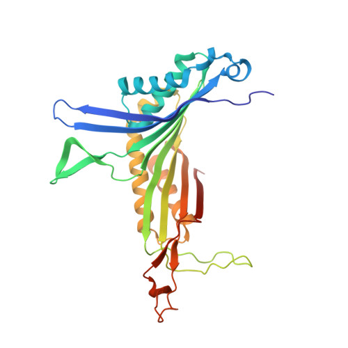
The neutron structure of urate oxidase resolves a long-standing mechanistic conundrum and reveals unexpected changes in protonation.

Oksanen, E.Blakeley, M.P.El-Hajji, M.Ryde, U.Budayova-Spano, M.

(2014) PLoS One 9: e86651-e86651

  • DOI: https://doi.org/10.1371/journal.pone.0086651
  • Primary Citation of Related Structures:  
    4N3M, 4N9M, 4N9S, 4N9V

  • PubMed Abstract: 

    Urate oxidase transforms uric acid to 5-hydroxyisourate without the help of cofactors, but the catalytic mechanism has remained enigmatic, as the protonation state of the substrate could not be reliably deduced. We have determined the neutron structure of urate oxidase, providing unique information on the proton positions. A neutron crystal structure inhibited by a chloride anion at 2.3 Å resolution shows that the substrate is in fact 8-hydroxyxanthine, the enol tautomer of urate. We have also determined the neutron structure of the complex with the inhibitor 8-azaxanthine at 1.9 Å resolution, showing the protonation states of the K10-T57-H256 catalytic triad. Together with X-ray data and quantum chemical calculations, these structures allow us to identify the site of the initial substrate protonation and elucidate why the enzyme is inhibited by a chloride anion.


  • Organizational Affiliation

    Institut de Biologie Structurale (IBS), Direction des Sciences du Vivant, Commissariat à l'Energie Atomique et aux Energies Alternatives, Grenoble, France, IBS, Centre National de la Recherche Scientifique, Grenoble, France, IBS, Université Grenoble Alpes, Grenoble, France.


Macromolecules
Find similar proteins by:  (by identity cutoff)  |  3D Structure
Entity ID: 1
MoleculeChains Sequence LengthOrganismDetailsImage
Uricase302Aspergillus flavusMutation(s): 0 
Gene Names: uaZuox
EC: 1.7.3.3
UniProt
Find proteins for Q00511 (Aspergillus flavus)
Explore Q00511 
Go to UniProtKB:  Q00511
Entity Groups  
Sequence Clusters30% Identity50% Identity70% Identity90% Identity95% Identity100% Identity
UniProt GroupQ00511
Sequence Annotations
Expand
  • Reference Sequence
Small Molecules
Ligands 3 Unique
IDChains Name / Formula / InChI Key2D Diagram3D Interactions
AZA
Query on AZA

Download Ideal Coordinates CCD File 
B [auth A]8-AZAXANTHINE
C4 H3 N5 O2
KVGVQTOQSNJTJI-UHFFFAOYSA-N
CL
Query on CL

Download Ideal Coordinates CCD File 
C [auth A]CHLORIDE ION
Cl
VEXZGXHMUGYJMC-UHFFFAOYSA-M
NA
Query on NA

Download Ideal Coordinates CCD File 
D [auth A]SODIUM ION
Na
FKNQFGJONOIPTF-UHFFFAOYSA-N
Experimental Data & Validation

Experimental Data

  • Method: NEUTRON DIFFRACTION
  • Resolution: 1.90 Å
  • R-Value Free: 0.267 
  • R-Value Work: 0.240 
  • R-Value Observed: 0.241 
  • Space Group: I 2 2 2
  • Method: X-RAY DIFFRACTION
  • Resolution: 1.92 Å
  • R-Value Free: 0.172 
  • R-Value Work: 0.140 
  • R-Value Observed: 0.141 
  • Space Group: I 2 2 2
Unit Cell:
Length ( Å )Angle ( ˚ )
a = 80.2α = 90
b = 96.2β = 90
c = 105.5γ = 90
Software Package:
Software NamePurpose
LADI-IIIdata collection
MOLREPphasing
PHENIXrefinement
LAUEGENdata reduction
SCALAdata scaling

Structure Validation

View Full Validation Report


Entry History 

Deposition Data

Revision History  (Full details and data files)

  • Version 1.0: 2014-02-05
    Type: Initial release
  • Version 1.1: 2014-02-26
    Changes: Database references
  • Version 1.2: 2018-06-13
    Changes: Data collection, Refinement description