4MZJ

Crystal Structure of MTIP from Plasmodium falciparum in complex with pGly[801,805], a stapled myoA tail peptide


Experimental Data Snapshot

  • Method: X-RAY DIFFRACTION
  • Resolution: 1.47 Å
  • R-Value Free: 0.216 
  • R-Value Work: 0.180 

wwPDB Validation   3D Report Full Report


This is version 1.5 of the entry. See complete history


Literature

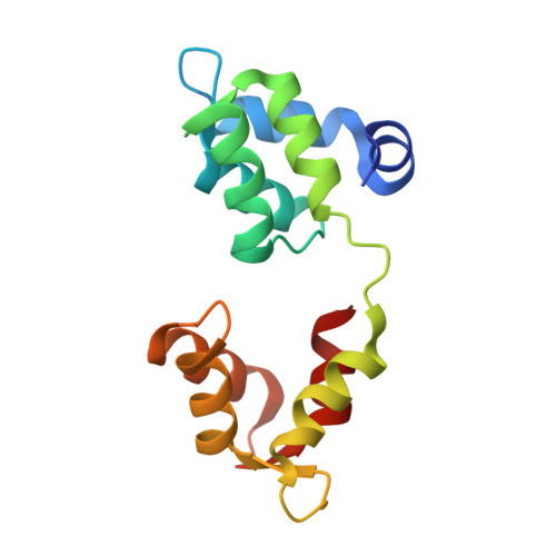
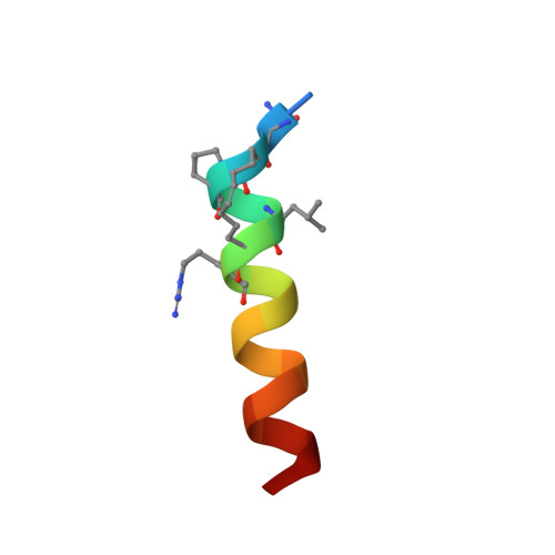
Crystal Structures of Stapled and Hydrogen Bond Surrogate Peptides Targeting a Fully Buried Protein-Helix Interaction.

Douse, C.H.Maas, S.J.Thomas, J.C.Garnett, J.A.Sun, Y.Cota, E.Tate, E.W.

(2014) ACS Chem Biol 8: 506-512

  • DOI: https://doi.org/10.1021/cb500271c
  • Primary Citation of Related Structures:  
    4MZJ, 4MZK, 4MZL

  • PubMed Abstract: 

    Constrained α-helical peptides are an exciting class of molecule designed to disrupt protein-protein interactions (PPIs) at a surface-exposed helix binding site. Complexes that engage more than one helical face account for over a third of structurally characterized helix PPIs, including several examples where the helix is fully buried. However, no constrained peptides have been reported that have targeted this class of interaction. We report the design of stapled and hydrogen bond surrogate (HBS) peptides mimicking the helical tail of the malaria parasite invasion motor myosin (myoA), which presents polar and hydrophobic functionality on all three faces in binding its partner, myoA tail interacting protein (MTIP), with high affinity. The first structures of these different constrained peptides bound to the same target are reported, enabling a direct comparison between these constraints and between staples based on monosubstituted pentenyl glycine (pGly) and disubstituted pentenyl alanine (pAla). Importantly, installation of these constraints does not disrupt native interactions in the buried site, so the affinity of the wild-type peptide is maintained.


  • Organizational Affiliation

    Department of Chemistry, ‡Centre for Structural Biology, Department of Life Sciences, and §Institute of Chemical Biology, Imperial College London , London SW7 2AZ, United Kingdom.


Macromolecules
Find similar proteins by:  (by identity cutoff)  |  3D Structure
Entity ID: 1
MoleculeChains Sequence LengthOrganismDetailsImage
Myosin A tail domain interacting protein145Plasmodium falciparum 3D7Mutation(s): 0 
Gene Names: MTIPPFL2225w
UniProt
Find proteins for Q8I4W8 (Plasmodium falciparum (isolate 3D7))
Explore Q8I4W8 
Go to UniProtKB:  Q8I4W8
Entity Groups  
Sequence Clusters30% Identity50% Identity70% Identity90% Identity95% Identity100% Identity
UniProt GroupQ8I4W8
Sequence Annotations
Expand
  • Reference Sequence

Find similar proteins by:  Sequence   |   3D Structure  

Entity ID: 2
MoleculeChains Sequence LengthOrganismDetailsImage
Myosin-AB [auth T]19Plasmodium falciparum 3D7Mutation(s): 3 
UniProt
Find proteins for Q8IDR3 (Plasmodium falciparum (isolate 3D7))
Explore Q8IDR3 
Go to UniProtKB:  Q8IDR3
Entity Groups  
Sequence Clusters30% Identity50% Identity70% Identity90% Identity95% Identity100% Identity
UniProt GroupQ8IDR3
Sequence Annotations
Expand
  • Reference Sequence
Small Molecules
Modified Residues  1 Unique
IDChains TypeFormula2D DiagramParent
NLE
Query on NLE
B [auth T]L-PEPTIDE LINKINGC6 H13 N O2LEU
Biologically Interesting Molecules (External Reference) 1 Unique
Experimental Data & Validation

Experimental Data

  • Method: X-RAY DIFFRACTION
  • Resolution: 1.47 Å
  • R-Value Free: 0.216 
  • R-Value Work: 0.180 
  • Space Group: P 21 21 21
Unit Cell:
Length ( Å )Angle ( ˚ )
a = 37.38α = 90
b = 53.98β = 90
c = 75.63γ = 90
Software Package:
Software NamePurpose
Adxvdata processing
PHASERphasing
PHENIXrefinement
XDSdata reduction
Aimlessdata scaling

Structure Validation

View Full Validation Report


Entry History 

Deposition Data

Revision History  (Full details and data files)

  • Version 1.0: 2013-11-06
    Type: Initial release
  • Version 1.1: 2014-08-20
    Changes: Database references
  • Version 1.2: 2014-09-03
    Changes: Database references
  • Version 1.3: 2017-11-15
    Changes: Refinement description
  • Version 1.4: 2023-09-20
    Changes: Data collection, Database references, Derived calculations, Refinement description
  • Version 1.5: 2023-12-06
    Changes: Data collection