4MYP

Structure of the central NEAT domain, N2, of the listerial Hbp2 protein complexed with heme


Experimental Data Snapshot

  • Method: X-RAY DIFFRACTION
  • Resolution: 1.80 Å
  • R-Value Free: 0.225 
  • R-Value Work: 0.195 
  • R-Value Observed: 0.196 

wwPDB Validation   3D Report Full Report


Ligand Structure Quality Assessment 


This is version 1.2 of the entry. See complete history


Literature

Novel Mechanism of Hemin Capture by Hbp2, the Hemoglobin-binding Hemophore from Listeria monocytogenes.

Malmirchegini, G.R.Sjodt, M.Shnitkind, S.Sawaya, M.R.Rosinski, J.Newton, S.M.Klebba, P.E.Clubb, R.T.

(2014) J Biol Chem 289: 34886-34899

  • DOI: https://doi.org/10.1074/jbc.M114.583013
  • Primary Citation of Related Structures:  
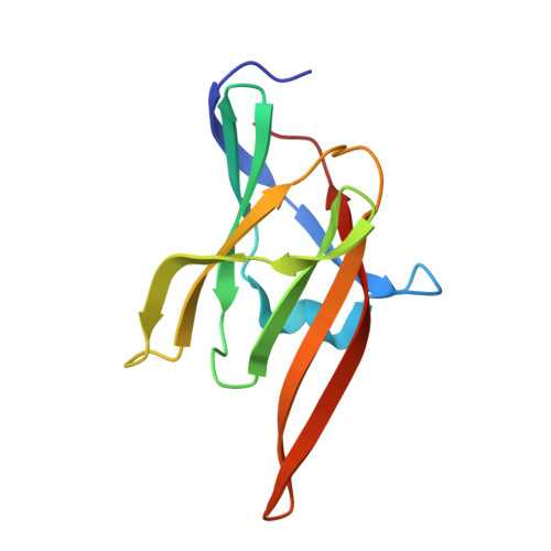
    4MYP, 4NLA

  • PubMed Abstract: 

    Iron is an essential nutrient that is required for the growth of the bacterial pathogen Listeria monocytogenes. In cell cultures, this microbe secretes hemin/hemoglobin-binding protein 2 (Hbp2; Lmo2185) protein, which has been proposed to function as a hemophore that scavenges heme from the environment. Based on its primary sequence, Hbp2 contains three NEAr transporter (NEAT) domains of unknown function. Here we show that each of these domains mediates high affinity binding to ferric heme (hemin) and that its N- and C-terminal domains interact with hemoglobin (Hb). The results of hemin transfer experiments are consistent with Hbp2 functioning as an Hb-binding hemophore that delivers hemin to other Hbp2 proteins that are attached to the cell wall. Surprisingly, our work reveals that the central NEAT domain in Hbp2 binds hemin even though its primary sequence lacks a highly conserved YXXXY motif that is used by all other previously characterized NEAT domains to coordinate iron in the hemin molecule. To elucidate the mechanism of hemin binding by Hbp2, we determined crystal structures of its central NEAT domain (Hbp2(N2); residues 183-303) in its free and hemin-bound states. The structures reveal an unprecedented mechanism of hemin binding in which Hbp2(N2) undergoes a major conformational rearrangement that facilitates metal coordination by a non-canonical tyrosine residue. These studies highlight previously unrecognized plasticity in the hemin binding mechanism of NEAT domains and provide insight into how L. monocytogenes captures heme iron.


  • Organizational Affiliation

    From the Department of Chemistry and Biochemistry and the UCLA-Department of Energy Institute for Genomics and Proteomics, UCLA, Los Angeles, California 90095 and.


Macromolecules
Find similar proteins by:  (by identity cutoff)  |  3D Structure
Entity ID: 1
MoleculeChains Sequence LengthOrganismDetailsImage
Iron-regulated surface determinant protein A
A, B
122Listeria monocytogenesMutation(s): 0 
Gene Names: BN418_2635lmo2185
UniProt
Find proteins for Q7AP54 (Listeria monocytogenes serovar 1/2a (strain ATCC BAA-679 / EGD-e))
Explore Q7AP54 
Go to UniProtKB:  Q7AP54
Entity Groups  
Sequence Clusters30% Identity50% Identity70% Identity90% Identity95% Identity100% Identity
UniProt GroupQ7AP54
Sequence Annotations
Expand
  • Reference Sequence
Small Molecules
Ligands 3 Unique
IDChains Name / Formula / InChI Key2D Diagram3D Interactions
HEM
Query on HEM

Download Ideal Coordinates CCD File 
C [auth A],
L [auth B]
PROTOPORPHYRIN IX CONTAINING FE
C34 H32 Fe N4 O4
KABFMIBPWCXCRK-RGGAHWMASA-L
GOL
Query on GOL

Download Ideal Coordinates CCD File 
G [auth A]
H [auth A]
I [auth A]
J [auth A]
K [auth A]
G [auth A],
H [auth A],
I [auth A],
J [auth A],
K [auth A],
N [auth B],
O [auth B],
P [auth B],
Q [auth B]
GLYCEROL
C3 H8 O3
PEDCQBHIVMGVHV-UHFFFAOYSA-N
ZN
Query on ZN

Download Ideal Coordinates CCD File 
D [auth A],
E [auth A],
F [auth A],
M [auth B]
ZINC ION
Zn
PTFCDOFLOPIGGS-UHFFFAOYSA-N
Experimental Data & Validation

Experimental Data

  • Method: X-RAY DIFFRACTION
  • Resolution: 1.80 Å
  • R-Value Free: 0.225 
  • R-Value Work: 0.195 
  • R-Value Observed: 0.196 
  • Space Group: P 1 21 1
Unit Cell:
Length ( Å )Angle ( ˚ )
a = 32.73α = 90
b = 67.69β = 91.62
c = 58.48γ = 90
Software Package:
Software NamePurpose
XSCALEdata scaling
PHASERphasing
BUSTER-TNTrefinement
PDB_EXTRACTdata extraction
XDSdata reduction
BUSTERrefinement

Structure Validation

View Full Validation Report


Ligand Structure Quality Assessment 


Entry History 

Deposition Data

Revision History  (Full details and data files)

  • Version 1.0: 2014-10-22
    Type: Initial release
  • Version 1.1: 2014-12-31
    Changes: Database references
  • Version 1.2: 2024-02-28
    Changes: Data collection, Database references, Derived calculations