4MN4

Structural Basis for the MukB-topoisomerase IV Interaction


Experimental Data Snapshot

  • Method: X-RAY DIFFRACTION
  • Resolution: 2.30 Å
  • R-Value Free: 0.246 
  • R-Value Work: 0.204 
  • R-Value Observed: 0.206 

wwPDB Validation   3D Report Full Report


This is version 1.3 of the entry. See complete history


Literature

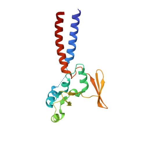
Structural basis for the MukB-topoisomerase IV interaction and its functional implications in vivo.

Vos, S.M.Stewart, N.K.Oakley, M.G.Berger, J.M.

(2013) EMBO J 32: 2950-2962

  • DOI: https://doi.org/10.1038/emboj.2013.218
  • Primary Citation of Related Structures:  
    4MN4

  • PubMed Abstract: 

    Chromosome partitioning in Escherichia coli is assisted by two interacting proteins, topoisomerase (topo) IV and MukB. MukB stimulates the relaxation of negative supercoils by topo IV; to understand the mechanism of their action and to define this functional interplay, we determined the crystal structure of a minimal MukB-topo IV complex to 2.3 Å resolution. The structure shows that the so-called 'hinge' region of MukB forms a heterotetrameric assembly with a C-terminal DNA binding domain (CTD) on topo IV's ParC subunit. Biochemical studies show that the hinge stimulates topo IV by competing for a site on the CTD that normally represses activity on negatively supercoiled DNA, while complementation tests using mutants implicated in the interaction reveal that the cellular dependency on topo IV derives from a joint need for both strand passage and MukB binding. Interestingly, the configuration of the MukB·topo IV complex sterically disfavours intradimeric interactions, indicating that the proteins may form oligomeric arrays with one another, and suggesting a framework by which MukB and topo IV may collaborate during daughter chromosome disentanglement.


  • Organizational Affiliation

    Department of Molecular and Cell Biology, University of California, Berkeley, CA, USA.


Macromolecules
Find similar proteins by:  (by identity cutoff)  |  3D Structure
Entity ID: 1
MoleculeChains Sequence LengthOrganismDetailsImage
DNA topoisomerase 4 subunit AA,
D [auth B]
259Escherichia coli str. K-12 substr. MG1655Mutation(s): 0 
Gene Names: parC
EC: 5.99.1.3
UniProt
Find proteins for P0AFI2 (Escherichia coli (strain K12))
Explore P0AFI2 
Go to UniProtKB:  P0AFI2
Entity Groups  
Sequence Clusters30% Identity50% Identity70% Identity90% Identity95% Identity100% Identity
UniProt GroupP0AFI2
Sequence Annotations
Expand
  • Reference Sequence
Find similar proteins by:  (by identity cutoff)  |  3D Structure
Entity ID: 2
MoleculeChains Sequence LengthOrganismDetailsImage
Chromosome partition protein MukBB [auth C],
C [auth D]
163Escherichia coli str. K-12 substr. MG1655Mutation(s): 0 
Gene Names: mukB
UniProt
Find proteins for P22523 (Escherichia coli (strain K12))
Explore P22523 
Go to UniProtKB:  P22523
Entity Groups  
Sequence Clusters30% Identity50% Identity70% Identity90% Identity95% Identity100% Identity
UniProt GroupP22523
Sequence Annotations
Expand
  • Reference Sequence
Small Molecules
Modified Residues  1 Unique
IDChains TypeFormula2D DiagramParent
MSE
Query on MSE
A,
D [auth B]
L-PEPTIDE LINKINGC5 H11 N O2 SeMET
Experimental Data & Validation

Experimental Data

  • Method: X-RAY DIFFRACTION
  • Resolution: 2.30 Å
  • R-Value Free: 0.246 
  • R-Value Work: 0.204 
  • R-Value Observed: 0.206 
  • Space Group: P 21 21 21
Unit Cell:
Length ( Å )Angle ( ˚ )
a = 49.767α = 90
b = 103.737β = 90
c = 186.611γ = 90
Software Package:
Software NamePurpose
Blu-Icedata collection
PHENIXmodel building
PHENIXrefinement
HKL-2000data reduction
HKL-2000data scaling
PHENIXphasing

Structure Validation

View Full Validation Report


Entry History 

Deposition Data

Revision History  (Full details and data files)

  • Version 1.0: 2013-10-23
    Type: Initial release
  • Version 1.1: 2014-01-22
    Changes: Database references
  • Version 1.2: 2023-09-20
    Changes: Data collection, Database references, Derived calculations, Refinement description
  • Version 1.3: 2023-12-06
    Changes: Data collection