4MM1

GGGPS from Methanothermobacter thermautotrophicus


Experimental Data Snapshot

  • Method: X-RAY DIFFRACTION
  • Resolution: 2.80 Å
  • R-Value Free: 0.271 
  • R-Value Work: 0.200 
  • R-Value Observed: 0.204 

wwPDB Validation   3D Report Full Report


Ligand Structure Quality Assessment 


This is version 1.3 of the entry. See complete history


Literature

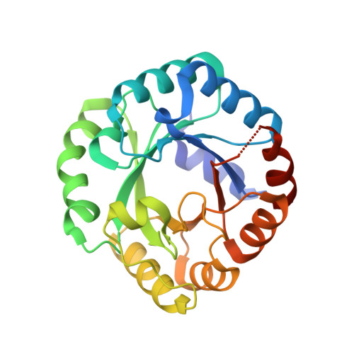
A comprehensive analysis of the geranylgeranylglyceryl phosphate synthase enzyme family identifies novel members and reveals mechanisms of substrate specificity and quaternary structure organization.

Peterhoff, D.Beer, B.Rajendran, C.Kumpula, E.P.Kapetaniou, E.Guldan, H.Wierenga, R.K.Sterner, R.Babinger, P.

(2014) Mol Microbiol 92: 885-899

  • DOI: https://doi.org/10.1111/mmi.12596
  • Primary Citation of Related Structures:  
    4JEJ, 4MM1, 4NAE, 4NAF

  • PubMed Abstract: 

    Geranylgeranylglyceryl phosphate synthase (GGGPS) family enzymes catalyse the formation of an ether bond between glycerol-1-phosphate and polyprenyl diphosphates. They are essential for the biosynthesis of archaeal membrane lipids, but also occur in bacterial species, albeit with unknown physiological function. It has been known that there exist two phylogenetic groups (I and II) of GGGPS family enzymes, but a comprehensive study has been missing. We therefore visualized the variability within the family by applying a sequence similarity network, and biochemically characterized 17 representative GGGPS family enzymes regarding their catalytic activities and substrate specificities. Moreover, we present the first crystal structures of group II archaeal and bacterial enzymes. Our analysis revealed that the previously uncharacterized bacterial enzymes from group II have GGGPS activity like the archaeal enzymes and differ from the bacterial group I enzymes that are heptaprenylglyceryl phosphate synthases. The length of the isoprenoid substrate is determined in group II GGGPS enzymes by 'limiter residues' that are different from those in group I enzymes, as shown by site-directed mutagenesis. Most of the group II enzymes form hexamers. We could disrupt these hexamers to stable and catalytically active dimers by mutating a single amino acid that acts as an 'aromatic anchor'.


  • Organizational Affiliation

    Institute of Biophysics and Physical Biochemistry, University of Regensburg, Regensburg, 93040, Germany.


Macromolecules
Find similar proteins by:  (by identity cutoff)  |  3D Structure
Entity ID: 1
MoleculeChains Sequence LengthOrganismDetailsImage
Geranylgeranylglyceryl phosphate synthase
A, B, C, D, E
A, B, C, D, E, F
250Methanothermobacter thermautotrophicus str. Delta HMutation(s): 0 
Gene Names: MTH_552
EC: 2.5.1.41
UniProt
Find proteins for O26652 (Methanothermobacter thermautotrophicus (strain ATCC 29096 / DSM 1053 / JCM 10044 / NBRC 100330 / Delta H))
Explore O26652 
Go to UniProtKB:  O26652
Entity Groups  
Sequence Clusters30% Identity50% Identity70% Identity90% Identity95% Identity100% Identity
UniProt GroupO26652
Sequence Annotations
Expand
  • Reference Sequence
Small Molecules
Ligands 2 Unique
IDChains Name / Formula / InChI Key2D Diagram3D Interactions
1GP
Query on 1GP

Download Ideal Coordinates CCD File 
G [auth A]
I [auth B]
K [auth C]
M [auth D]
O [auth E]
G [auth A],
I [auth B],
K [auth C],
M [auth D],
O [auth E],
Q [auth F]
SN-GLYCEROL-1-PHOSPHATE
C3 H9 O6 P
AWUCVROLDVIAJX-VKHMYHEASA-N
PGE
Query on PGE

Download Ideal Coordinates CCD File 
H [auth A]
J [auth B]
L [auth C]
N [auth D]
P [auth E]
H [auth A],
J [auth B],
L [auth C],
N [auth D],
P [auth E],
R [auth F]
TRIETHYLENE GLYCOL
C6 H14 O4
ZIBGPFATKBEMQZ-UHFFFAOYSA-N
Experimental Data & Validation

Experimental Data

  • Method: X-RAY DIFFRACTION
  • Resolution: 2.80 Å
  • R-Value Free: 0.271 
  • R-Value Work: 0.200 
  • R-Value Observed: 0.204 
  • Space Group: C 1 2 1
Unit Cell:
Length ( Å )Angle ( ˚ )
a = 143.675α = 90
b = 148.756β = 109.5
c = 91.503γ = 90
Software Package:
Software NamePurpose
PHENIXmodel building
PHENIXrefinement
XDSdata reduction
XDSdata scaling
PHENIXphasing

Structure Validation

View Full Validation Report


Ligand Structure Quality Assessment 


Entry History 

Deposition Data

Revision History  (Full details and data files)

  • Version 1.0: 2014-06-25
    Type: Initial release
  • Version 1.1: 2017-11-15
    Changes: Refinement description
  • Version 1.2: 2024-02-28
    Changes: Data collection, Database references, Derived calculations
  • Version 1.3: 2024-03-13
    Changes: Source and taxonomy