4M7R

Crystal structure of the N-terminal methyltransferase-like domain of anamorsin


Experimental Data Snapshot

  • Method: X-RAY DIFFRACTION
  • Resolution: 1.80 Å
  • R-Value Free: 0.226 
  • R-Value Work: 0.187 

wwPDB Validation   3D Report Full Report


This is version 1.1 of the entry. See complete history


Literature

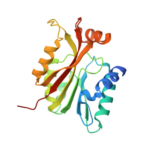
Crystal structure of the N-terminal methyltransferase-like domain of anamorsin.

Song, G.Cheng, C.Li, Y.Shaw, N.Xiao, Z.C.Liu, Z.J.

(2014) Proteins 82: 1066-1071

  • DOI: https://doi.org/10.1002/prot.24443
  • Primary Citation of Related Structures:  
    4M7R

  • PubMed Abstract: 

    Anamorsin is a recently identified molecule that inhibits apoptosis during hematopoiesis. It contains an N-terminal methyltransferase-like domain and a C-terminal Fe-S cluster motif. Not much is known about the function of the protein. To better understand the function of anamorsin, we have solved the crystal structure of the N-terminal domain at 1.8 Å resolution. Although the overall structure resembles a typical S-adenosylmethionine (SAM) dependent methyltransferase fold, it lacks one α-helix and one β-strand. As a result, the N-terminal domain as well as the full-length anamorsin did not show S-adenosyl-L-methionine (AdoMet) dependent methyltransferase activity. Structural comparisons with known AdoMet dependent methyltransferases reveals subtle differences in the SAM binding pocket that preclude the N-terminal domain from binding to AdoMet. The N-terminal methyltransferase-like domain of anamorsin probably functions as a structural scaffold to inhibit methyl transfers by out-competing other AdoMet dependant methyltransferases or acts as bait for protein-protein interactions.


  • Organizational Affiliation

    National Laboratory of Biomacromolecules, Institute of Biophysics, Chinese Academy of Sciences, Beijing, 100101, China.


Macromolecules
Find similar proteins by:  (by identity cutoff)  |  3D Structure
Entity ID: 1
MoleculeChains Sequence LengthOrganismDetailsImage
Anamorsin
A, B
196Homo sapiensMutation(s): 0 
Gene Names: CIAPIN1CUA001PRO0915
UniProt & NIH Common Fund Data Resources
Find proteins for Q6FI81 (Homo sapiens)
Explore Q6FI81 
Go to UniProtKB:  Q6FI81
PHAROS:  Q6FI81
GTEx:  ENSG00000005194 
Entity Groups  
Sequence Clusters30% Identity50% Identity70% Identity90% Identity95% Identity100% Identity
UniProt GroupQ6FI81
Sequence Annotations
Expand
  • Reference Sequence
Small Molecules
Ligands 1 Unique
IDChains Name / Formula / InChI Key2D Diagram3D Interactions
HG
Query on HG

Download Ideal Coordinates CCD File 
C [auth A]MERCURY (II) ION
Hg
BQPIGGFYSBELGY-UHFFFAOYSA-N
Experimental Data & Validation

Experimental Data

  • Method: X-RAY DIFFRACTION
  • Resolution: 1.80 Å
  • R-Value Free: 0.226 
  • R-Value Work: 0.187 
  • Space Group: P 61
Unit Cell:
Length ( Å )Angle ( ˚ )
a = 79.508α = 90
b = 79.508β = 90
c = 101.75γ = 120
Software Package:
Software NamePurpose
HKL-2000data collection
SOLVEphasing
PHENIXrefinement
HKL-2000data reduction
HKL-2000data scaling

Structure Validation

View Full Validation Report


Entry History 

Deposition Data

Revision History  (Full details and data files)

  • Version 1.0: 2013-10-30
    Type: Initial release
  • Version 1.1: 2022-08-24
    Changes: Database references, Derived calculations