4LZF

A novel domain in the microcephaly protein CPAP suggests a role in centriole architecture


Experimental Data Snapshot

  • Method: X-RAY DIFFRACTION
  • Resolution: 1.72 Å
  • R-Value Free: 0.231 
  • R-Value Work: 0.190 
  • R-Value Observed: 0.192 

wwPDB Validation   3D Report Full Report


This is version 1.4 of the entry. See complete history


Literature

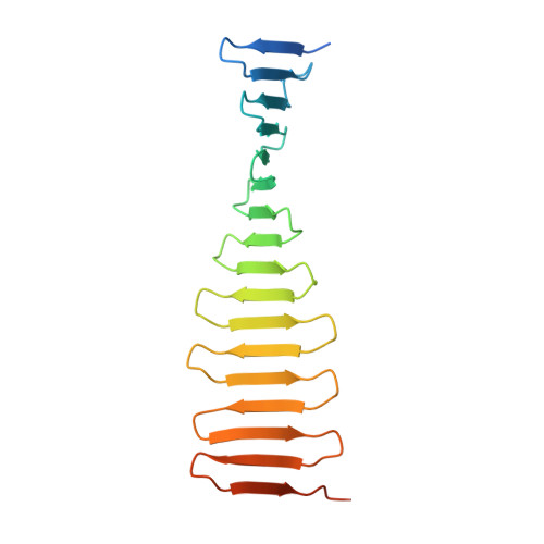
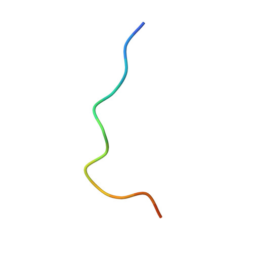
Structural analysis of the G-box domain of the microcephaly protein CPAP suggests a role in centriole architecture.

Hatzopoulos, G.N.Erat, M.C.Cutts, E.Rogala, K.B.Slater, L.M.Stansfeld, P.J.Vakonakis, I.

(2013) Structure 21: 2069-2077

  • DOI: https://doi.org/10.1016/j.str.2013.08.019
  • Primary Citation of Related Structures:  
    4LD1, 4LD3, 4LZF

  • PubMed Abstract: 

    Centrioles are evolutionarily conserved eukaryotic organelles composed of a protein scaffold surrounded by sets of microtubules organized with a 9-fold radial symmetry. CPAP, a centriolar protein essential for microtubule recruitment, features a C-terminal domain of unknown structure, the G-box. A missense mutation in the G-box reduces affinity for the centriolar shuttling protein STIL and causes primary microcephaly. Here, we characterize the molecular architecture of CPAP and determine the G-box structure alone and in complex with a STIL fragment. The G-box comprises a single elongated β sheet capable of forming supramolecular assemblies. Structural and biophysical studies highlight the conserved nature of the CPAP-STIL complex. We propose that CPAP acts as a horizontal "strut" that joins the centriolar scaffold with microtubules, whereas G-box domains form perpendicular connections.


  • Organizational Affiliation

    Department of Biochemistry, University of Oxford, South Parks Road, Oxford OX1 3QU, UK.


Macromolecules
Find similar proteins by:  (by identity cutoff)  |  3D Structure
Entity ID: 1
MoleculeChains Sequence LengthOrganismDetailsImage
Centrosomal P4.1-associated protein184Danio rerioMutation(s): 0 
Gene Names: cenpj
UniProt
Find proteins for E7FCY1 (Danio rerio)
Explore E7FCY1 
Go to UniProtKB:  E7FCY1
Entity Groups  
Sequence Clusters30% Identity50% Identity70% Identity90% Identity95% Identity100% Identity
UniProt GroupE7FCY1
Sequence Annotations
Expand
  • Reference Sequence

Find similar proteins by:  Sequence   |   3D Structure  

Entity ID: 2
MoleculeChains Sequence LengthOrganismDetailsImage
SCL-interrupting locus protein homolog15Danio rerioMutation(s): 0 
UniProt
Find proteins for Q8JGS1 (Danio rerio)
Explore Q8JGS1 
Go to UniProtKB:  Q8JGS1
Entity Groups  
Sequence Clusters30% Identity50% Identity70% Identity90% Identity95% Identity100% Identity
UniProt GroupQ8JGS1
Sequence Annotations
Expand
  • Reference Sequence
Experimental Data & Validation

Experimental Data

  • Method: X-RAY DIFFRACTION
  • Resolution: 1.72 Å
  • R-Value Free: 0.231 
  • R-Value Work: 0.190 
  • R-Value Observed: 0.192 
  • Space Group: C 1 2 1
Unit Cell:
Length ( Å )Angle ( ˚ )
a = 97.58α = 90
b = 38.39β = 126.68
c = 70.05γ = 90
Software Package:
Software NamePurpose
Aimlessdata scaling
PHASERphasing
PHENIXrefinement
PDB_EXTRACTdata extraction
XDSdata scaling
XDSdata reduction

Structure Validation

View Full Validation Report


Entry History 

Deposition Data

Revision History  (Full details and data files)

  • Version 1.0: 2013-09-11
    Type: Initial release
  • Version 1.1: 2013-10-16
    Changes: Database references
  • Version 1.2: 2014-02-05
    Changes: Database references
  • Version 1.3: 2018-01-24
    Changes: Structure summary
  • Version 1.4: 2023-09-20
    Changes: Data collection, Database references, Derived calculations, Refinement description