4LY4

Crystal structure of peptidoglycan deacetylase (HP0310) with Zinc from Helicobacter pylori


Experimental Data Snapshot

  • Method: X-RAY DIFFRACTION
  • Resolution: 2.20 Å
  • R-Value Free: 0.207 
  • R-Value Work: 0.169 
  • R-Value Observed: 0.171 

wwPDB Validation   3D Report Full Report


This is version 1.4 of the entry. See complete history


Literature

Characterization of the divalent metal binding site of bacterial polysaccharide deacetylase using crystallography and quantum chemical calculations.

Shaik, M.M.Bhattacharjee, N.Bhattacharjee, A.Field, M.J.Zanotti, G.

(2014) Proteins 82: 1311-1318

  • DOI: https://doi.org/10.1002/prot.24497
  • Primary Citation of Related Structures:  
    4LY4

  • PubMed Abstract: 

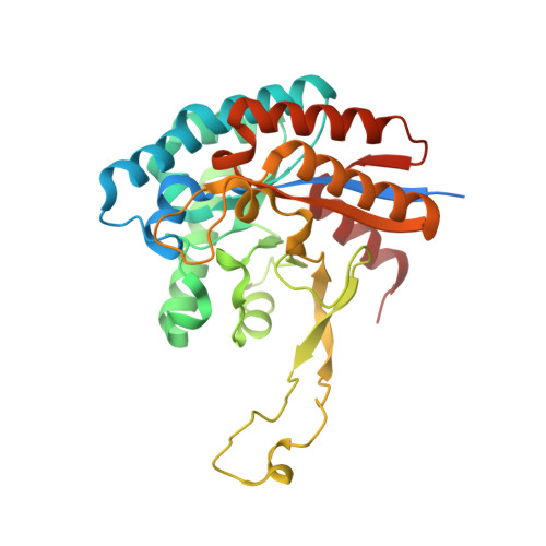
    Peptidoglycan deacetlyase (HP0310, HpPgdA) from the gram-negative pathogen Helicobacter pylori, is the enzyme responsible for a peptidoglycan modification that counteracts the host immune response. In a recent study, we determined the crystallographic structure of the enzyme, which is a homo-tetramer (Shaik et al., PloS One 2011;6:e19207). The metal-binding site, which is essential for the enzyme's catalytic activity, is visible within the structure, but we were unable to identify the nature of the metal itself. In this study, we have obtained a higher-resolution crystal structure of the enzyme, which shows that the ion bound is, in fact, zinc. Analysis of the structure of the four sites, one per monomer, and quantum chemical calculations of models of the site in the presence of different divalent metal ions show an intrinsic preference for zinc, but also significant flexibility of the site so that binding of other ions can eventually occur.


  • Organizational Affiliation

    PATBAC, Institut de Biologie Structurale-Jean-Pierre Ebel, Grenoble, France.


Macromolecules
Find similar proteins by:  (by identity cutoff)  |  3D Structure
Entity ID: 1
MoleculeChains Sequence LengthOrganismDetailsImage
peptidoglycan deacetylase
A, B, C, D
326Helicobacter pylori G27Mutation(s): 0 
Gene Names: HP0310HPG27_289
UniProt
Find proteins for B5ZA76 (Helicobacter pylori (strain G27))
Explore B5ZA76 
Go to UniProtKB:  B5ZA76
Entity Groups  
Sequence Clusters30% Identity50% Identity70% Identity90% Identity95% Identity100% Identity
UniProt GroupB5ZA76
Sequence Annotations
Expand
  • Reference Sequence
Experimental Data & Validation

Experimental Data

  • Method: X-RAY DIFFRACTION
  • Resolution: 2.20 Å
  • R-Value Free: 0.207 
  • R-Value Work: 0.169 
  • R-Value Observed: 0.171 
  • Space Group: I 2 2 2
Unit Cell:
Length ( Å )Angle ( ˚ )
a = 113.64α = 90
b = 153.93β = 90
c = 153.88γ = 90
Software Package:
Software NamePurpose
ADSCdata collection
PHASERphasing
PHENIXrefinement
MOSFLMdata reduction
SCALAdata scaling

Structure Validation

View Full Validation Report


Entry History 

Deposition Data

Revision History  (Full details and data files)

  • Version 1.0: 2013-08-28
    Type: Initial release
  • Version 1.1: 2013-11-20
    Changes: Structure summary
  • Version 1.2: 2014-01-22
    Changes: Database references
  • Version 1.3: 2014-10-08
    Changes: Database references
  • Version 1.4: 2023-09-20
    Changes: Data collection, Database references, Derived calculations, Refinement description