4LQ6

Crystal structure of Rv3717 reveals a novel amidase from M. tuberculosis


Experimental Data Snapshot

  • Method: X-RAY DIFFRACTION
  • Resolution: 1.68 Å
  • R-Value Free: 0.184 
  • R-Value Work: 0.161 
  • R-Value Observed: 0.162 

wwPDB Validation   3D Report Full Report


This is version 1.1 of the entry. See complete history


Literature

The structure of Rv3717 reveals a novel amidase from Mycobacterium tuberculosis.

Kumar, A.Kumar, S.Kumar, D.Mishra, A.Dewangan, R.P.Shrivastava, P.Ramachandran, S.Taneja, B.

(2013) Acta Crystallogr D Biol Crystallogr 69: 2543-2554

  • DOI: https://doi.org/10.1107/S0907444913026371
  • Primary Citation of Related Structures:  
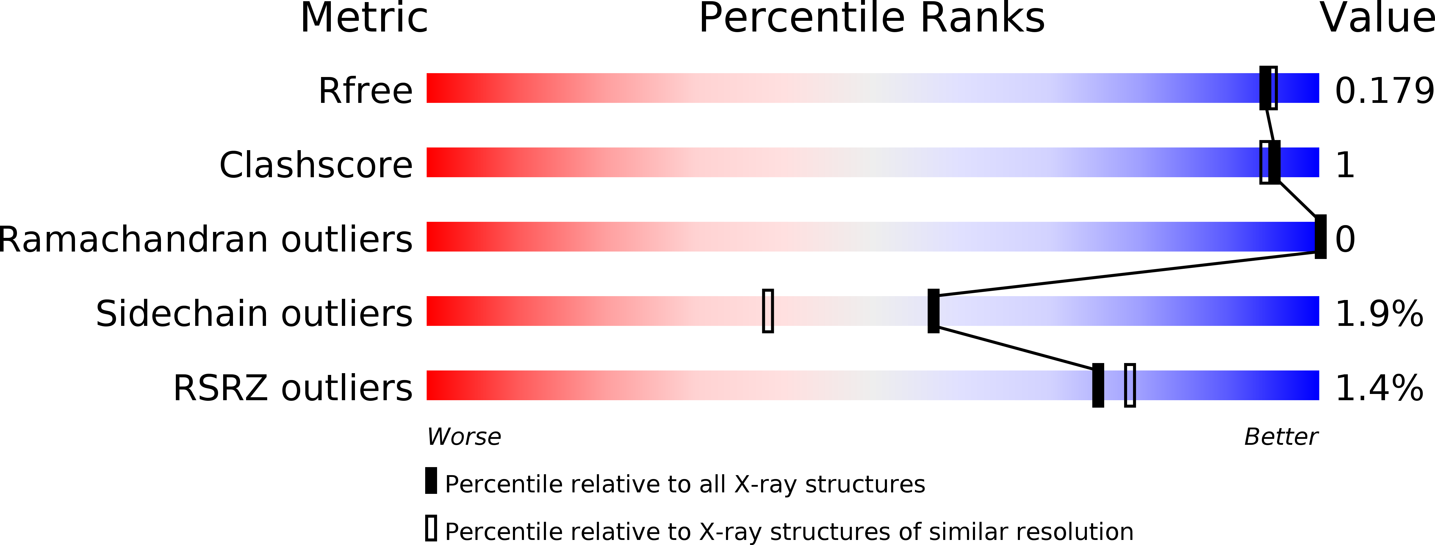
    4LQ6

  • PubMed Abstract: 

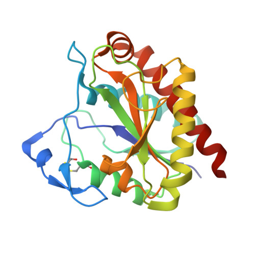
    Bacterial N-acetylmuramoyl-L-alanine amidases are cell-wall hydrolases that hydrolyze the bond between N-acetylmuramic acid and L-alanine in cell-wall glycopeptides. Rv3717 of Mycobacterium tuberculosis has been identified as a unique autolysin that lacks a cell-wall-binding domain (CBD) and its structure has been determined to 1.7 Å resolution by the Pt-SAD phasing method. Rv3717 possesses an α/β-fold and is a zinc-dependent hydrolase. The structure reveals a short flexible hairpin turn that partially occludes the active site and may be involved in autoregulation. This type of autoregulation of activity of PG hydrolases has been observed in Bartonella henselae amidase (AmiB) and may be a general mechanism used by some of the redundant amidases to regulate cell-wall hydrolase activity in bacteria. Rv3717 utilizes its net positive charge for substrate binding and exhibits activity towards a broad spectrum of substrate cell walls. The enzymatic activity of Rv3717 was confirmed by isolation and identification of its enzymatic products by LC/MS. These studies indicate that Rv3717, an N-acetylmuramoyl-L-alanine amidase from M. tuberculosis, represents a new family of lytic amidases that do not have a separate CBD and are regulated conformationally.


  • Organizational Affiliation

    Structural Biology Unit, CSIR-IGIB, South Campus, Mathura Road, New Delhi 110 025, India.


Macromolecules
Find similar proteins by:  (by identity cutoff)  |  3D Structure
Entity ID: 1
MoleculeChains Sequence LengthOrganismDetailsImage
N-acetymuramyl-L-alanine amidase-related protein218Mycobacterium tuberculosisMutation(s): 0 
Gene Names: MT3820Rv3717
UniProt
Find proteins for O69684 (Mycobacterium tuberculosis (strain CDC 1551 / Oshkosh))
Explore O69684 
Go to UniProtKB:  O69684
Entity Groups  
Sequence Clusters30% Identity50% Identity70% Identity90% Identity95% Identity100% Identity
UniProt GroupO69684
Sequence Annotations
Expand
  • Reference Sequence
Small Molecules
Ligands 4 Unique
IDChains Name / Formula / InChI Key2D Diagram3D Interactions
PT
Query on PT

Download Ideal Coordinates CCD File 
H [auth A],
I [auth A],
J [auth A]
PLATINUM (II) ION
Pt
HRGDZIGMBDGFTC-UHFFFAOYSA-N
SO4
Query on SO4

Download Ideal Coordinates CCD File 
C [auth A]SULFATE ION
O4 S
QAOWNCQODCNURD-UHFFFAOYSA-L
ZN
Query on ZN

Download Ideal Coordinates CCD File 
B [auth A]ZINC ION
Zn
PTFCDOFLOPIGGS-UHFFFAOYSA-N
CL
Query on CL

Download Ideal Coordinates CCD File 
D [auth A],
E [auth A],
F [auth A],
G [auth A]
CHLORIDE ION
Cl
VEXZGXHMUGYJMC-UHFFFAOYSA-M
Experimental Data & Validation

Experimental Data

  • Method: X-RAY DIFFRACTION
  • Resolution: 1.68 Å
  • R-Value Free: 0.184 
  • R-Value Work: 0.161 
  • R-Value Observed: 0.162 
  • Space Group: I 21 21 21
Unit Cell:
Length ( Å )Angle ( ˚ )
a = 37.35α = 90
b = 103.78β = 90
c = 129.97γ = 90
Software Package:
Software NamePurpose
HKL-2000data collection
Auto-Rickshawphasing
BUSTERrefinement
MOSFLMdata reduction
SCALAdata scaling

Structure Validation

View Full Validation Report


Entry History 

Deposition Data

Revision History  (Full details and data files)

  • Version 1.0: 2013-12-04
    Type: Initial release
  • Version 1.1: 2014-01-15
    Changes: Database references