Functional and Structural Characterization of Vibrio cholerae Extracellular Serine Protease B, VesB.
Gadwal, S., Korotkov, K.V., Delarosa, J.R., Hol, W.G., Sandkvist, M.(2014) J Biol Chem 289: 8288-8298
- PubMed: 24459146
- DOI: https://doi.org/10.1074/jbc.M113.525261
- Primary Citation of Related Structures:
4LK4 - PubMed Abstract:
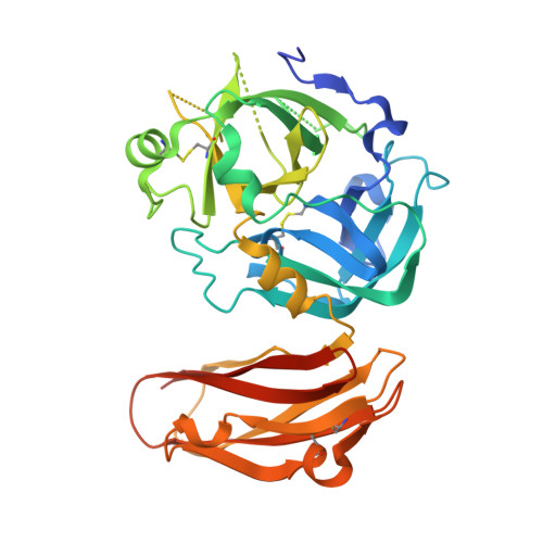
The chymotrypsin subfamily A of serine proteases consists primarily of eukaryotic proteases, including only a few proteases of bacterial origin. VesB, a newly identified serine protease that is secreted by the type II secretion system in Vibrio cholerae, belongs to this subfamily. VesB is likely produced as a zymogen because sequence alignment with trypsinogen identified a putative cleavage site for activation and a catalytic triad, His-Asp-Ser. Using synthetic peptides, VesB efficiently cleaved a trypsin substrate, but not chymotrypsin and elastase substrates. The reversible serine protease inhibitor, benzamidine, inhibited VesB and served as an immobilized ligand for VesB affinity purification, further indicating its relationship with trypsin-like enzymes. Consistent with this family of serine proteases, N-terminal sequencing implied that the propeptide is removed in the secreted form of VesB. Separate mutagenesis of the activation site and catalytic serine rendered VesB inactive, confirming the importance of these features for activity, but not for secretion. Similar to trypsin but, in contrast to thrombin and other coagulation factors, Na(+) did not stimulate the activity of VesB, despite containing the Tyr(250) signature. The crystal structure of catalytically inactive pro-VesB revealed that the protease domain is structurally similar to trypsinogen. The C-terminal domain of VesB was found to adopt an immunoglobulin (Ig)-fold that is structurally homologous to Ig-folds of other extracellular Vibrio proteins. Possible roles of the Ig-fold domain in stability, substrate specificity, cell surface association, and type II secretion of VesB, the first bacterial multidomain trypsin-like protease with known structure, are discussed.
Organizational Affiliation:
From the Department of Microbiology and Immunology, University of Michigan Medical School, Ann Arbor, Michigan 48109 and.