4LFV

Crystal structure of human FPPS in complex with YS0470 and two molecules of inorganic phosphate


Experimental Data Snapshot

  • Method: X-RAY DIFFRACTION
  • Resolution: 2.00 Å
  • R-Value Free: 0.218 
  • R-Value Work: 0.176 
  • R-Value Observed: 0.179 

wwPDB Validation   3D Report Full Report


Ligand Structure Quality Assessment 


This is version 1.2 of the entry. See complete history


Literature

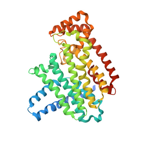
Structure of human farnesyl pyrophosphate synthase in complex with an aminopyridine bisphosphonate and two molecules of inorganic phosphate.

Park, J.Lin, Y.S.Tsantrizos, Y.S.Berghuis, A.M.

(2014) Acta Crystallogr Sect F Struct Biol Cryst Commun 70: 299-304

  • DOI: https://doi.org/10.1107/S2053230X14002106
  • Primary Citation of Related Structures:  
    4LFV

  • PubMed Abstract: 

    Human farnesyl pyrophosphate synthase (hFPPS) produces farnesyl pyrophosphate, an isoprenoid essential for a variety of cellular processes. The enzyme has been well established as the molecular target of the nitrogen-containing bisphosphonates (N-BPs), which are best known for their antiresorptive effects in bone but are also known for their anticancer properties. Crystal structures of hFPPS in ternary complexes with a novel bisphosphonate, YS0470, and the secondary ligands inorganic phosphate (Pi), inorganic pyrophosphate (PPi) and isopentenyl pyrophosphate (IPP) have recently been reported. Only the co-binding of the bisphosphonate with either PPi or IPP resulted in the full closure of the C-terminal tail of the enzyme, a conformational change that is required for catalysis and that is also responsible for the potent in vivo efficacy of N-BPs. In the present communication, a co-crystal structure of hFPPS in complex with YS0470 and two molecules of Pi is reported. The unusually close proximity between these ligands, which was confirmed by anomalous diffraction data, suggests that they interact with one another, with their anionic charges neutralized in their bound state. The structure also showed the tail of the enzyme to be fully disordered, indicating that simultaneous binding of two Pi molecules with a bisphosphonate cannot induce the tail-closing conformational change in hFPPS. Examination of homologous FPPSs suggested that this ligand-dependent tail closure is only conserved in the mammalian proteins. The prevalence of Pi-bound hFPPS structures in the PDB raises a question regarding the in vivo relevance of Pi binding to the function of the enzyme.


  • Organizational Affiliation

    Department of Biochemistry, McGill University, 3655 Promenade Sir William Osler, Montreal, QC H3G 1Y6, Canada.


Macromolecules
Find similar proteins by:  (by identity cutoff)  |  3D Structure
Entity ID: 1
MoleculeChains Sequence LengthOrganismDetailsImage
Farnesyl pyrophosphate synthaseA [auth F]375Homo sapiensMutation(s): 0 
Gene Names: FDPSFPSKIAA1293
EC: 2.5.1.10 (PDB Primary Data), 2.5.1.1 (PDB Primary Data)
UniProt & NIH Common Fund Data Resources
Find proteins for P14324 (Homo sapiens)
Explore P14324 
Go to UniProtKB:  P14324
PHAROS:  P14324
GTEx:  ENSG00000160752 
Entity Groups  
Sequence Clusters30% Identity50% Identity70% Identity90% Identity95% Identity100% Identity
UniProt GroupP14324
Sequence Annotations
Expand
  • Reference Sequence
Small Molecules
Ligands 4 Unique
IDChains Name / Formula / InChI Key2D Diagram3D Interactions
YS4
Query on YS4

Download Ideal Coordinates CCD File 
B [auth F][({4-[4-(propan-2-yloxy)phenyl]pyridin-2-yl}amino)methanediyl]bis(phosphonic acid)
C15 H20 N2 O7 P2
DTXBFSJBRGLOHO-UHFFFAOYSA-N
PO4
Query on PO4

Download Ideal Coordinates CCD File 
C [auth F],
D [auth F]
PHOSPHATE ION
O4 P
NBIIXXVUZAFLBC-UHFFFAOYSA-K
CL
Query on CL

Download Ideal Coordinates CCD File 
H [auth F]CHLORIDE ION
Cl
VEXZGXHMUGYJMC-UHFFFAOYSA-M
MG
Query on MG

Download Ideal Coordinates CCD File 
E [auth F],
F,
G [auth F]
MAGNESIUM ION
Mg
JLVVSXFLKOJNIY-UHFFFAOYSA-N
Binding Affinity Annotations 
IDSourceBinding Affinity
YS4 BindingDB:  4LFV IC50: min: 28, max: 1390 (nM) from 2 assay(s)
Experimental Data & Validation

Experimental Data

  • Method: X-RAY DIFFRACTION
  • Resolution: 2.00 Å
  • R-Value Free: 0.218 
  • R-Value Work: 0.176 
  • R-Value Observed: 0.179 
  • Space Group: P 41 21 2
Unit Cell:
Length ( Å )Angle ( ˚ )
a = 111.02α = 90
b = 111.02β = 90
c = 67.04γ = 90
Software Package:
Software NamePurpose
MxDCdata collection
REFMACrefinement
XDSdata reduction
XSCALEdata scaling
REFMACphasing

Structure Validation

View Full Validation Report


Ligand Structure Quality Assessment 


Entry History 

Deposition Data

Revision History  (Full details and data files)

  • Version 1.0: 2014-03-12
    Type: Initial release
  • Version 1.1: 2014-08-27
    Changes: Database references
  • Version 1.2: 2023-09-20
    Changes: Data collection, Database references, Derived calculations, Refinement description