4L4J

Crystal structure of fc-fragment of human IgG2-Sigma antibody


Experimental Data Snapshot

  • Method: X-RAY DIFFRACTION
  • Resolution: 1.92 Å
  • R-Value Free: 0.247 
  • R-Value Work: 0.200 
  • R-Value Observed: 0.201 

wwPDB Validation   3D Report Full Report


This is version 2.1 of the entry. See complete history


Literature

An engineered Fc variant of an IgG eliminates all immune effector functions via structural perturbations.

Vafa, O.Gilliland, G.L.Brezski, R.J.Strake, B.Wilkinson, T.Lacy, E.R.Scallon, B.Teplyakov, A.Malia, T.J.Strohl, W.R.

(2014) Methods 65: 114-126

  • DOI: https://doi.org/10.1016/j.ymeth.2013.06.035
  • Primary Citation of Related Structures:  
    4L4J

  • PubMed Abstract: 

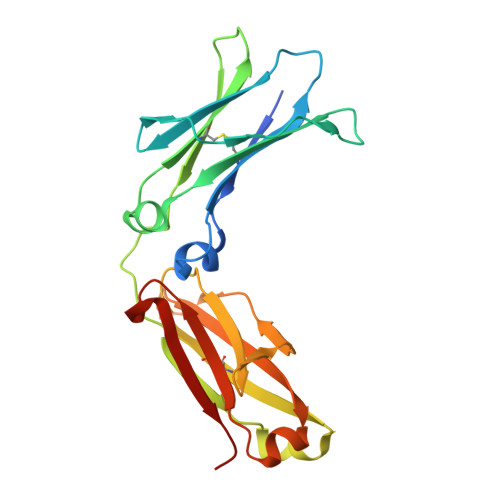
    The Fc variant of IgG2, designated as IgG2σ, was engineered with V234A/G237A /P238S/H268A/V309L/A330S/P331S substitutions to eliminate affinity for Fcγ receptors and C1q complement protein and consequently, immune effector functions. IgG2σ was compared to other previously well-characterized Fc 'muted' variants, including aglycosylated IgG1, IgG2m4 (H268Q/V309L/A330S/P331S, changes to IgG4), and IgG4 ProAlaAla (S228P/L234A/L235A) in its capacity to bind FcγRs and activate various immune-stimulatory responses. In contrast to the previously characterized muted Fc variants, which retain selective FcγR binding and effector functions, IgG2σ shows no detectable binding to the Fcγ receptors in affinity and avidity measurements, nor any detectable antibody-dependent cytotoxicity, phagocytosis, complement activity, or Fc-mediated cytokine release. Moreover, IgG2σ shows minimal immunogenic potential by T-cell epitope analysis. The circulating half-life of IgG2σ in monkeys is extended relative to IgG1 and IgG2, in spite of similar in vitro binding to recombinant FcRn. The three-dimensional structure of the Fc, needed for assessing the basis for the absence of effector function, was compared with that of IgG2 revealing a number of conformational differences near the hinge region of the CH2 domain that result from the amino acid substitutions. Modeling reveals that at least one of the key interactions with FcγRs is disrupted by a conformational change that reorients P329 to a position that prevents it from interacting with conserved W90 and W113 residues of the FcγRs. Inspection of the structure also indicated significant changes to the conformations of D270 and P329 in the CH2 domain that could negatively impact C1q binding. Thus, structural perturbations of the Fc provide a rationale for the loss of function. In toto, these properties of IgG2σ suggest that it is a superior alternative to previously described IgG variants of minimal effector function, for future therapeutic applications of non-immunostimulatory mAb and Fc-fusion platforms.


  • Organizational Affiliation

    Biologics Research, Biotechnology Center of Excellence, Janssen Research & Development, LLC, 1400 McKean Road, Spring House, PA 19477, United States. Electronic address: ovafa@its.jnj.com.


Macromolecules
Find similar proteins by:  (by identity cutoff)  |  3D Structure
Entity ID: 1
MoleculeChains Sequence LengthOrganismDetailsImage
Ig gamma-2 chain C region
A, B
221Homo sapiensMutation(s): 7 
Gene Names: IGHG2
UniProt & NIH Common Fund Data Resources
Find proteins for P01859 (Homo sapiens)
Explore P01859 
Go to UniProtKB:  P01859
PHAROS:  P01859
GTEx:  ENSG00000211893 
Entity Groups  
Sequence Clusters30% Identity50% Identity70% Identity90% Identity95% Identity100% Identity
UniProt GroupP01859
Sequence Annotations
Expand
  • Reference Sequence
Oligosaccharides

Help

Entity ID: 2
MoleculeChains Length2D Diagram Glycosylation3D Interactions
2-acetamido-2-deoxy-beta-D-glucopyranose-(1-2)-alpha-D-mannopyranose-(1-6)-[alpha-D-mannopyranose-(1-3)]beta-D-mannopyranose-(1-4)-2-acetamido-2-deoxy-beta-D-glucopyranose-(1-4)-[alpha-L-fucopyranose-(1-6)]2-acetamido-2-deoxy-beta-D-glucopyranose
C
7N-Glycosylation
Glycosylation Resources
GlyTouCan:  G45889JQ
GlyCosmos:  G45889JQ
GlyGen:  G45889JQ
Entity ID: 3
MoleculeChains Length2D Diagram Glycosylation3D Interactions
2-acetamido-2-deoxy-beta-D-glucopyranose-(1-2)-alpha-D-mannopyranose-(1-6)-beta-D-mannopyranose-(1-4)-2-acetamido-2-deoxy-beta-D-glucopyranose-(1-4)-2-acetamido-2-deoxy-beta-D-glucopyranose
D
5N/A
Glycosylation Resources
GlyTouCan:  G12045WP
GlyCosmos:  G12045WP
Small Molecules
Ligands 1 Unique
IDChains Name / Formula / InChI Key2D Diagram3D Interactions
GOL
Query on GOL

Download Ideal Coordinates CCD File 
E [auth A]GLYCEROL
C3 H8 O3
PEDCQBHIVMGVHV-UHFFFAOYSA-N
Experimental Data & Validation

Experimental Data

  • Method: X-RAY DIFFRACTION
  • Resolution: 1.92 Å
  • R-Value Free: 0.247 
  • R-Value Work: 0.200 
  • R-Value Observed: 0.201 
  • Space Group: P 21 21 21
Unit Cell:
Length ( Å )Angle ( ˚ )
a = 68.54α = 90
b = 69.97β = 90
c = 112.25γ = 90
Software Package:
Software NamePurpose
StructureStudiodata collection
PHASERphasing
REFMACrefinement
XDSdata reduction
XDSdata scaling

Structure Validation

View Full Validation Report


Entry History 

Deposition Data

Revision History  (Full details and data files)

  • Version 1.0: 2013-08-28
    Type: Initial release
  • Version 1.1: 2014-01-22
    Changes: Database references
  • Version 2.0: 2020-07-29
    Type: Remediation
    Reason: Carbohydrate remediation
    Changes: Advisory, Atomic model, Data collection, Database references, Derived calculations, Structure summary
  • Version 2.1: 2023-09-20
    Changes: Data collection, Database references, Refinement description, Structure summary