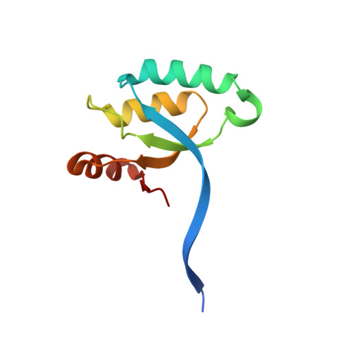
Crystal structure of the N-terminal domain of MinC dimerized via domain swapping.
An, J.Y., Kim, T.G., Park, K.R., Lee, J.G., Youn, H.S., Lee, Y., Kang, J.Y., Kang, G.B., Eom, S.H.(2013) J Synchrotron Radiat 20: 984-988
- PubMed: 24121353
- DOI: https://doi.org/10.1107/S0909049513022760
- Primary Citation of Related Structures:
4L1C - PubMed Abstract:
Proper cell division at the mid-site of gram-negative bacteria reflects critical regulation by the min system (MinC, MinD and MinE) of the cytokinetic Z ring, which is a polymer composed of FtsZ subunits. MinC and MinD act together to inhibit aberrantly positioned Z-ring formation. MinC consists of two domains: an N-terminal domain (MinCNTD), which interacts with FtsZ and inhibits FtsZ polymerization, and a C-terminal domain (MinCCTD), which interacts with MinD and inhibits the bundling of FtsZ filaments. These two domains reportedly function together, and both are essential for normal cell division. The full-length dimeric structure of MinC from Thermotoga maritima has been reported, and shows that MinC dimerization occurs via MinCCTD; MinCNTD is not involved in dimerization. Here the crystal structure of Escherichia coli MinCNTD (EcoMinCNTD) is reported. EcoMinCNTD forms a dimer via domain swapping between the first β strands in each subunit. It is therefore suggested that the dimerization of full-length EcoMinC occurs via both MinCCTD and MinCNTD, and that the dimerized EcoMinCNTD likely plays an important role in inhibiting aberrant Z-ring localization.
Organizational Affiliation:
School of Life Sciences, Gwangju Institute of Science and Technology, Gwangju 500-712, Republic of Korea.