4KRU

X-ray structure of catalytic domain of endolysin from clostridium perfringens phage phiSM101


Experimental Data Snapshot

  • Method: X-RAY DIFFRACTION
  • Resolution: 1.37 Å
  • R-Value Free: 0.193 
  • R-Value Work: 0.172 
  • R-Value Observed: 0.172 

wwPDB Validation   3D Report Full Report


Ligand Structure Quality Assessment 


This is version 2.1 of the entry. See complete history


Literature

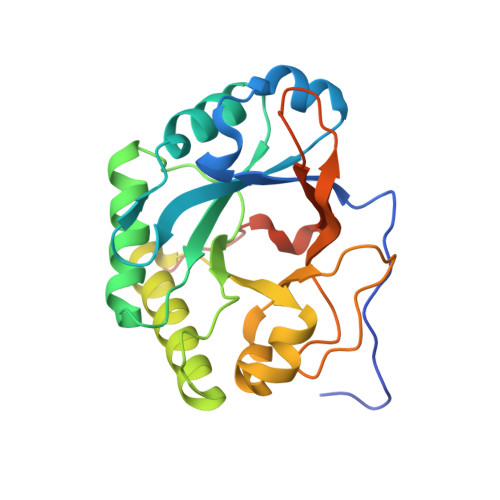
X-ray structure of a novel endolysin encoded by episomal phage phiSM101 of Clostridium perfringens.

Tamai, E.Yoshida, H.Sekiya, H.Nariya, H.Miyata, S.Okabe, A.Kuwahara, T.Maki, J.Kamitori, S.

(2014) Mol Microbiol 92: 326-337

  • DOI: https://doi.org/10.1111/mmi.12559
  • Primary Citation of Related Structures:  
    4KRT, 4KRU

  • PubMed Abstract: 

    Gram-positive bacteria possess a thick cell wall composed of a mesh polymer of peptidoglycans, which provides physical protection. Endolysins encoded by phages infecting bacteria can hydrolyse peptidoglycans in the bacterial cell wall, killing the host bacteria immediately. The endolysin (Psm) encoded by episomal phage phiSM101 of enterotoxigenic Clostridium perfringens type A strain SM101 exhibits potent lytic activity towards most strains of Clostridium perfringens. Psm has an N-terminal catalytic domain highly homologous to N-acetylmuramidases belonging to the glycoside hydrolase 25 family, and C-terminal tandem repeated bacterial Src homology 3 (SH3_3) domains as the cell wall-binding domain. The X-ray structure of full-length Psm and a catalytic domain of Psm in complex with N-acetylglucosamine were determined to elucidate the catalytic reaction and cell wall recognition mechanisms of Psm. The results showed that Psm may have adopted a neighbouring-group mechanism for the catalytic hydrolysing reaction in which the N-acetyl carbonyl group of the substrate was involved in the formation of an oxazolinium ion intermediate. Based on structural comparisons with other endolysins and a modelling study, we proposed that tandem repeated SH3_3 domains of Psm recognized the peptide side-chains of peptidoglycans to assist the catalytic domain hydrolysing the glycan backbone.


  • Organizational Affiliation

    Life Science Research Center, Kagawa University, 1750-1, Ikenobe, Miki-cho, Kita-gun, Kagawa, 761-0793, Japan; Department of Infectious Disease, College of Pharmaceutical Science, Matsuyama University, 4-2 Bunkyo-cho, Matsuyama, Ehime, 790-8578, Japan.


Macromolecules
Find similar proteins by:  (by identity cutoff)  |  3D Structure
Entity ID: 1
MoleculeChains Sequence LengthOrganismDetailsImage
Autolytic lysozyme229Clostridium phage phiSM101Mutation(s): 0 
Gene Names: CPR_C0050
EC: 3.2.1.17
UniProt
Find proteins for Q0SPG7 (Clostridium phage phiSM101)
Explore Q0SPG7 
Go to UniProtKB:  Q0SPG7
Entity Groups  
Sequence Clusters30% Identity50% Identity70% Identity90% Identity95% Identity100% Identity
UniProt GroupQ0SPG7
Sequence Annotations
Expand
  • Reference Sequence
Small Molecules
Ligands 3 Unique
IDChains Name / Formula / InChI Key2D Diagram3D Interactions
NAG
Query on NAG

Download Ideal Coordinates CCD File 
C [auth A]2-acetamido-2-deoxy-beta-D-glucopyranose
C8 H15 N O6
OVRNDRQMDRJTHS-FMDGEEDCSA-N
NDG
Query on NDG

Download Ideal Coordinates CCD File 
B [auth A]2-acetamido-2-deoxy-alpha-D-glucopyranose
C8 H15 N O6
OVRNDRQMDRJTHS-PVFLNQBWSA-N
PO4
Query on PO4

Download Ideal Coordinates CCD File 
D [auth A]PHOSPHATE ION
O4 P
NBIIXXVUZAFLBC-UHFFFAOYSA-K
Experimental Data & Validation

Experimental Data

  • Method: X-RAY DIFFRACTION
  • Resolution: 1.37 Å
  • R-Value Free: 0.193 
  • R-Value Work: 0.172 
  • R-Value Observed: 0.172 
  • Space Group: P 21 21 21
Unit Cell:
Length ( Å )Angle ( ˚ )
a = 47.77α = 90
b = 57.62β = 90
c = 73.42γ = 90
Software Package:
Software NamePurpose
CrystalCleardata collection
MOLREPphasing
CNSrefinement
CrystalCleardata reduction
CrystalCleardata scaling

Structure Validation

View Full Validation Report


Ligand Structure Quality Assessment 


Entry History 

Deposition Data

Revision History  (Full details and data files)

  • Version 1.0: 2014-04-02
    Type: Initial release
  • Version 1.1: 2019-12-18
    Changes: Database references
  • Version 2.0: 2020-07-29
    Type: Remediation
    Reason: Carbohydrate remediation
    Changes: Atomic model, Data collection, Derived calculations, Structure summary
  • Version 2.1: 2023-11-08
    Changes: Data collection, Database references, Refinement description, Structure summary