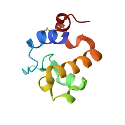
Carbohydrate Recognition by RpfB from Mycobacterium tuberculosis Unveiled by Crystallographic and Molecular Dynamics Analyses.
Squeglia, F., Romano, M., Ruggiero, A., Vitagliano, L., De Simone, A., Berisio, R.(2013) Biophys J 104: 2530-2539
- PubMed: 23746526
- DOI: https://doi.org/10.1016/j.bpj.2013.04.040
- Primary Citation of Related Structures:
4KL7, 4KPM - PubMed Abstract:
Resuscitation of Mtb is crucial to the etiology of Tuberculosis, because latent tuberculosis is estimated to affect one-third of the world population. The resuscitation-promoting factor RpfB is mainly responsible for Mtb resuscitation from dormancy. Given the impact of latent Tuberculosis, RpfB represents an interesting target for tuberculosis drug discovery. However, no molecular models of substrate binding and catalysis are hitherto available for this enzyme. Here, we identified key interactions involved in substrate binding to RpfB by combining x-ray diffraction studies and computational approaches. The crystal structure of RpfB catalytic domain in complex with N,N',N"-triacetyl-chitotriose, as described here, provides the first, to our knowledge, atomic representation of ligand recognition by RpfB and demonstrates that the strongest interactions are established by the N-acetylglucosamine moiety in the central region of the enzyme binding cleft. Molecular dynamics analyses provided information on the dynamic behavior of protein-substrate interactions and on the role played by the solvent in RpfB function. These data combined with sequence conservation analysis suggest that Glu-292 is the sole residue crucial for catalysis, implying that RpfB acts via the formation of an oxocarbenium ion rather than a covalent intermediate. Present data represent a solid base for the design of effective drug inhibitors of RpfB. Moreover, homology models were generated for the catalytic domains of all members of the Mtb Rpf family (RpfA-E). The analysis of these models unveiled analogies and differences among the different members of the Rpf protein family.
Organizational Affiliation:
Institute of Biostructures and Bioimaging, C.N.R., Naples, Italy.