


Crystal structure of catalytic domain of endolysin Ply40
Romero-Fernandez, P., Bartual, S.G., Carrasco-lopez, C., Loessner, M., Hermoso, J.A.To be published.
Experimental Data Snapshot
wwPDB Validation 3D Report Full Report

Entity ID: 1 | |||||
|---|---|---|---|---|---|
| Molecule | Chains | Sequence Length | Organism | Details | Image |
| Gp26 | 223 | Listeria phage P40 | Mutation(s): 0 Gene Names: gp26 | 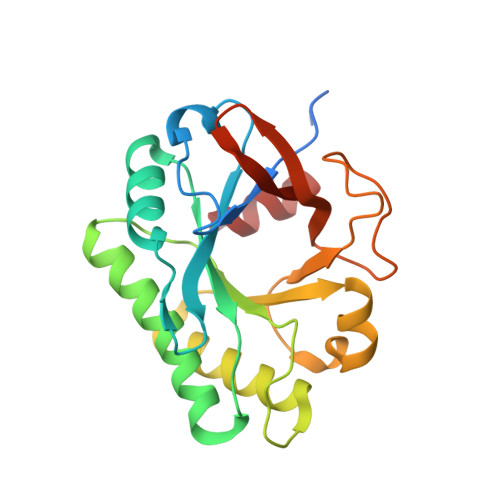 | |
UniProt | |||||
Find proteins for B6D7J9 (Listeria phage P40) Explore B6D7J9 Go to UniProtKB: B6D7J9 | |||||
Entity Groups | |||||
| Sequence Clusters | 30% Identity50% Identity70% Identity90% Identity95% Identity100% Identity | ||||
| UniProt Group | B6D7J9 | ||||
Sequence AnnotationsExpand | |||||
| |||||
| Length ( Å ) | Angle ( ˚ ) |
|---|---|
| a = 42.332 | α = 90 |
| b = 42.332 | β = 90 |
| c = 98.627 | γ = 120 |
| Software Name | Purpose |
|---|---|
| ADSC | data collection |
| PHASER | phasing |
| PHENIX | refinement |
| XDS | data reduction |
| Aimless | data scaling |
RCSB PDB (citation) is hosted by
RCSB PDB is a member of the

