


X-ray Crystal Structure of a Putative Lipoprotein from Bacillus anthracis
Brunzelle, J.S., Wawrzak, Z., Onopriyenko, O., Anderson, W.F., Savchenko, A., Center for Structural Genomics of Infectious DiseasesTo be published.
Experimental Data Snapshot
wwPDB Validation 3D Report Full Report

Entity ID: 1 | |||||
|---|---|---|---|---|---|
| Molecule | Chains | Sequence Length | Organism | Details | Image |
| Lipoprotein | 174 | Bacillus anthracis | Mutation(s): 0 Gene Names: BA_0580, BAS0549, GBAA_0580 | 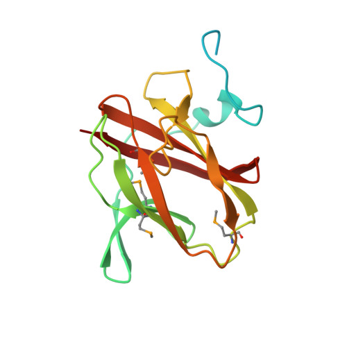 | |
UniProt | |||||
Find proteins for A0A6L7HR41 (Bacillus anthracis) Explore A0A6L7HR41 Go to UniProtKB: A0A6L7HR41 | |||||
Entity Groups | |||||
| Sequence Clusters | 30% Identity50% Identity70% Identity90% Identity95% Identity100% Identity | ||||
| UniProt Group | A0A6L7HR41 | ||||
Sequence AnnotationsExpand | |||||
| |||||
| Modified Residues 1 Unique | |||||
|---|---|---|---|---|---|
| ID | Chains | Type | Formula | 2D Diagram | Parent |
| MSE Query on MSE | A, B | L-PEPTIDE LINKING | C5 H11 N O2 Se |  | MET |
| Length ( Å ) | Angle ( ˚ ) |
|---|---|
| a = 85.863 | α = 90 |
| b = 85.863 | β = 90 |
| c = 236.078 | γ = 120 |
| Software Name | Purpose |
|---|---|
| BLU-MAX | data collection |
| PHENIX | model building |
| PHENIX | refinement |
| XDS | data reduction |
| Aimless | data scaling |
| PHENIX | phasing |
RCSB PDB (citation) is hosted by
RCSB PDB is a member of the

