4JFA

Crystal Structure of Plasmodium falciparum Tryptophanyl-tRNA synthetase


Experimental Data Snapshot

  • Method: X-RAY DIFFRACTION
  • Resolution: 2.60 Å
  • R-Value Free: 0.229 
  • R-Value Work: 0.191 
  • R-Value Observed: 0.192 

wwPDB Validation   3D Report Full Report


Ligand Structure Quality Assessment 


This is version 1.0 of the entry. See complete history


Literature

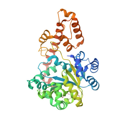
An appended domain results in an unusual architecture for malaria parasite tryptophanyl-tRNA synthetase

Khan, S.Garg, A.Sharma, A.Camacho, N.Picchioni, D.Saint-Leger, A.Ribas de Pouplana, L.Yogavel, M.Sharma, A.

(2013) PLoS One 8: e66224-e66224

  • DOI: https://doi.org/10.1371/journal.pone.0066224
  • Primary Citation of Related Structures:  
    4JFA

  • PubMed Abstract: 

    Specific activation of amino acids by aminoacyl-tRNA synthetases (aaRSs) is essential for maintaining fidelity during protein translation. Here, we present crystal structure of malaria parasite Plasmodium falciparum tryptophanyl-tRNA synthetase (Pf-WRS) catalytic domain (AAD) at 2.6 Å resolution in complex with L-tryptophan. Confocal microscopy-based localization data suggest cytoplasmic residency of this protein. Pf-WRS has an unusual N-terminal extension of AlaX-like domain (AXD) along with linker regions which together seem vital for enzymatic activity and tRNA binding. Pf-WRS is not proteolytically processed in the parasites and therefore AXD likely provides tRNA binding capability rather than editing activity. The N-terminal domain containing AXD and linker region is monomeric and would result in an unusual overall architecture for Pf-WRS where the dimeric catalytic domains have monomeric AXDs on either side. Our PDB-wide comparative analyses of 47 WRS crystal structures also provide new mechanistic insights into this enzyme family in context conserved KMSKS loop conformations.


  • Organizational Affiliation

    Structural and Computational Biology Group, International Centre for Genetic Engineering and Biotechnology (ICGEB), New Delhi, India.


Macromolecules
Find similar proteins by:  (by identity cutoff)  |  3D Structure
Entity ID: 1
MoleculeChains Sequence LengthOrganismDetailsImage
Tryptophan--tRNA ligase
A, B, C, D
420Plasmodium falciparum 3D7Mutation(s): 0 
Gene Names: PF13_0205
EC: 6.1.1.2
UniProt
Find proteins for Q8IDW3 (Plasmodium falciparum (isolate 3D7))
Explore Q8IDW3 
Go to UniProtKB:  Q8IDW3
Entity Groups  
Sequence Clusters30% Identity50% Identity70% Identity90% Identity95% Identity100% Identity
UniProt GroupQ8IDW3
Sequence Annotations
Expand
  • Reference Sequence
Small Molecules
Ligands 3 Unique
IDChains Name / Formula / InChI Key2D Diagram3D Interactions
TRP
Query on TRP

Download Ideal Coordinates CCD File 
E [auth A],
G [auth B],
I [auth C],
K [auth D]
TRYPTOPHAN
C11 H12 N2 O2
QIVBCDIJIAJPQS-VIFPVBQESA-N
BME
Query on BME

Download Ideal Coordinates CCD File 
F [auth A],
H [auth B],
L [auth D]
BETA-MERCAPTOETHANOL
C2 H6 O S
DGVVWUTYPXICAM-UHFFFAOYSA-N
K
Query on K

Download Ideal Coordinates CCD File 
J [auth C],
M [auth D]
POTASSIUM ION
K
NPYPAHLBTDXSSS-UHFFFAOYSA-N
Experimental Data & Validation

Experimental Data

  • Method: X-RAY DIFFRACTION
  • Resolution: 2.60 Å
  • R-Value Free: 0.229 
  • R-Value Work: 0.191 
  • R-Value Observed: 0.192 
  • Space Group: P 1 21 1
Unit Cell:
Length ( Å )Angle ( ˚ )
a = 51.176α = 90
b = 213.252β = 97.15
c = 101.952γ = 90
Software Package:
Software NamePurpose
PHENIXrefinement
PDB_EXTRACTdata extraction
DNAdata collection
HKL-2000data reduction
HKL-2000data scaling
MOLREPphasing

Structure Validation

View Full Validation Report


Ligand Structure Quality Assessment 


Entry History 

Deposition Data

Revision History  (Full details and data files)

  • Version 1.0: 2014-01-29
    Type: Initial release