Crystal and NMR structures give insights into the role and dynamics of subunit F of the eukaryotic V-ATPase from Saccharomyces cerevisiae
Basak, S., Lim, J., Manimekalai, M.S.S., Balakrishna, A.M., Gruber, G.(2013) J Biol Chem 288: 11930-11939
- PubMed: 23476018
- DOI: https://doi.org/10.1074/jbc.M113.461533
- Primary Citation of Related Structures:
4IX9 - PubMed Abstract:
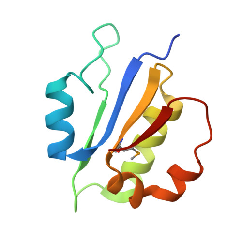
Subunit F of V-ATPases is proposed to undergo structural alterations during catalysis and reversible dissociation from the V1VO complex. Recently, we determined the low resolution structure of F from Saccharomyces cerevisiae V-ATPase, showing an N-terminal egg shape, connected to a C-terminal hook-like segment via a linker region. To understand the mechanistic role of subunit F of S. cerevisiae V-ATPase, composed of 118 amino acids, the crystal structure of the major part of F, F(1-94), was solved at 2.3 Å resolution. The structural features were confirmed by solution NMR spectroscopy using the entire F subunit. The eukaryotic F subunit consists of the N-terminal F(1-94) domain with four-parallel β-strands, which are intermittently surrounded by four α-helices, and the C terminus, including the α5-helix encompassing residues 103 to 113. Two loops (26)GQITPETQEK(35) and (60)ERDDI(64) are described to be essential in mechanistic processes of the V-ATPase enzyme. The (26)GQITPETQEK(35) loop becomes exposed when fitted into the recently determined EM structure of the yeast V1VO-ATPase. A mechanism is proposed in which the (26)GQITPETQEK(35) loop of subunit F and the flexible C-terminal domain of subunit H move in proximity, leading to an inhibitory effect of ATPase activity in V1. Subunits D and F are demonstrated to interact with subunit d. Together with NMR dynamics, the role of subunit F has been discussed in the light of its interactions in the processes of reversible disassembly and ATP hydrolysis of V-ATPases by transmitting movements of subunit d and H of the VO and V1 sector, respectively.
Organizational Affiliation:
Nanyang Technological University, School of Biological Sciences, 60 Nanyang Drive, Singapore 637551, Singapore.