4ITL

Crystal structure of LpxK from Aquifex aeolicus in complex with AMP-PCP at 2.1 angstrom resolution


Experimental Data Snapshot

  • Method: X-RAY DIFFRACTION
  • Resolution: 2.09 Å
  • R-Value Free: 0.190 
  • R-Value Work: 0.167 
  • R-Value Observed: 0.169 

wwPDB Validation   3D Report Full Report


Ligand Structure Quality Assessment 


This is version 1.3 of the entry. See complete history


Literature

Mechanistic Characterization of the Tetraacyldisaccharide-1-phosphate 4'-Kinase LpxK Involved in Lipid A Biosynthesis.

Emptage, R.P.Pemble, C.W.York, J.D.Raetz, C.R.Zhou, P.

(2013) Biochemistry 52: 2280-2290

  • DOI: https://doi.org/10.1021/bi400097z
  • Primary Citation of Related Structures:  
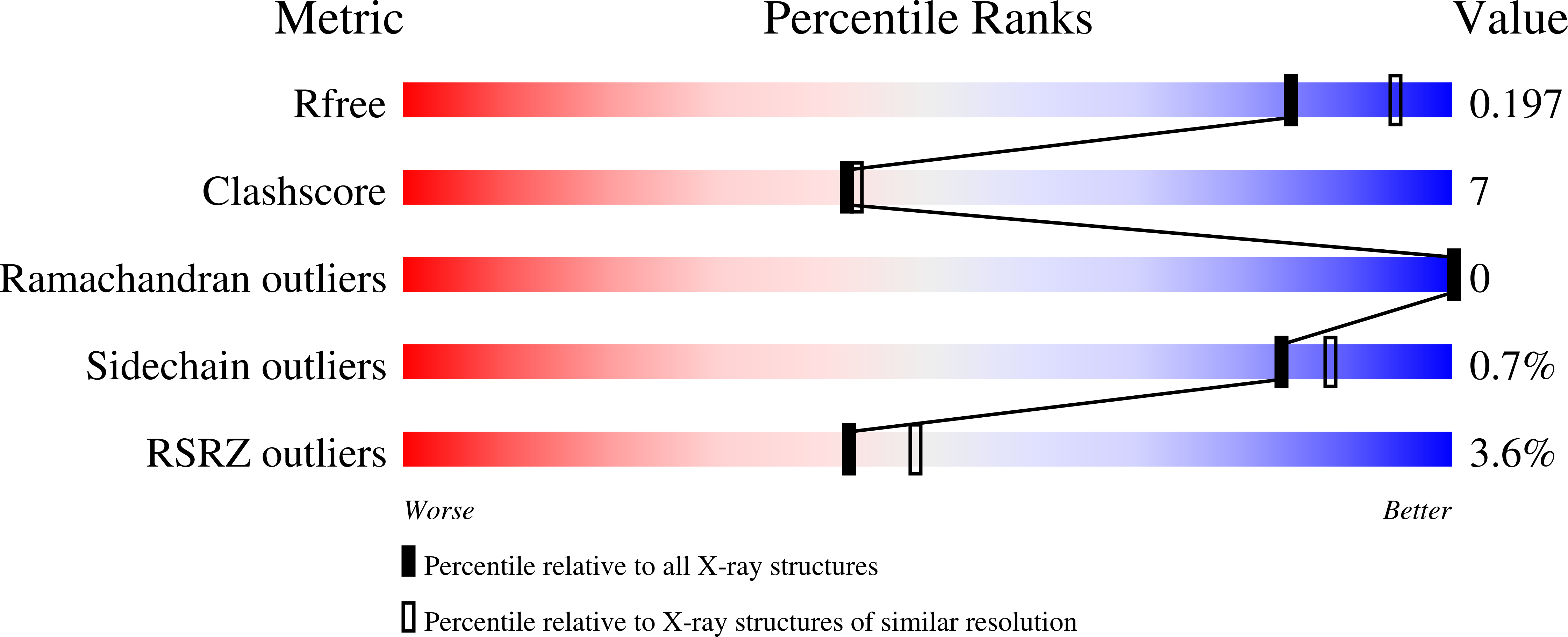
    4ITL, 4ITM, 4ITN

  • PubMed Abstract: 

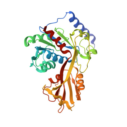
    The sixth step in the lipid A biosynthetic pathway involves phosphorylation of the tetraacyldisaccharide-1-phosphate (DSMP) intermediate by the cytosol-facing inner membrane kinase LpxK, a member of the P-loop-containing nucleoside triphosphate (NTP) hydrolase superfamily. We report the kinetic characterization of LpxK from Aquifex aeolicus and the crystal structures of LpxK in complex with ATP in a precatalytic binding state, the ATP analogue AMP-PCP in the closed catalytically competent conformation, and a chloride anion revealing an inhibitory conformation of the nucleotide-binding P-loop. We demonstrate that LpxK activity in vitro requires the presence of a detergent micelle and formation of a ternary LpxK-ATP/Mg(2+)-DSMP complex. Using steady-state kinetics, we have identified crucial active site residues, leading to the proposal that the interaction of D99 with H261 acts to increase the pKa of the imidazole moiety, which in turn serves as the catalytic base to deprotonate the 4'-hydroxyl of the DSMP substrate. The fact that an analogous mechanism has not yet been observed for other P-loop kinases highlights LpxK as a distinct member of the P-loop kinase family, a notion that is also reflected through its localization at the membrane, lipid substrate, and overall structure.


  • Organizational Affiliation

    Department of Biochemistry, Duke University Medical Center, Durham, NC 27710, USA. ryan.emptage@duke.edu


Macromolecules
Find similar proteins by:  (by identity cutoff)  |  3D Structure
Entity ID: 1
MoleculeChains Sequence LengthOrganismDetailsImage
Tetraacyldisaccharide 4'-kinase315Aquifex aeolicus VF5Mutation(s): 0 
Gene Names: lpxKaq_1656
EC: 2.7.1.130
UniProt
Find proteins for O67572 (Aquifex aeolicus (strain VF5))
Explore O67572 
Go to UniProtKB:  O67572
Entity Groups  
Sequence Clusters30% Identity50% Identity70% Identity90% Identity95% Identity100% Identity
UniProt GroupO67572
Sequence Annotations
Expand
  • Reference Sequence
Experimental Data & Validation

Experimental Data

  • Method: X-RAY DIFFRACTION
  • Resolution: 2.09 Å
  • R-Value Free: 0.190 
  • R-Value Work: 0.167 
  • R-Value Observed: 0.169 
  • Space Group: P 21 21 21
Unit Cell:
Length ( Å )Angle ( ˚ )
a = 66.086α = 90
b = 75.372β = 90
c = 104.56γ = 90
Software Package:
Software NamePurpose
DENZOdata reduction
SCALEPACKdata scaling
PHASERphasing
PHENIXrefinement
PDB_EXTRACTdata extraction
SERGUIdata collection
HKL-2000data reduction
HKL-2000data scaling

Structure Validation

View Full Validation Report


Ligand Structure Quality Assessment 


Entry History 

Deposition Data

Revision History  (Full details and data files)

  • Version 1.0: 2013-04-03
    Type: Initial release
  • Version 1.1: 2013-04-17
    Changes: Database references
  • Version 1.2: 2017-11-15
    Changes: Refinement description
  • Version 1.3: 2023-09-20
    Changes: Data collection, Database references, Derived calculations, Refinement description