4IJM

Crystal structure of circadian clock protein KaiC A422V mutant


Experimental Data Snapshot

  • Method: X-RAY DIFFRACTION
  • Resolution: 3.35 Å
  • R-Value Free: 0.261 
  • R-Value Work: 0.217 
  • R-Value Observed: 0.219 

wwPDB Validation   3D Report Full Report


Ligand Structure Quality Assessment 


This is version 1.3 of the entry. See complete history


Literature

Loop-Loop Interactions Regulate KaiA-Stimulated KaiC Phosphorylation in the Cyanobacterial KaiABC Circadian Clock.

Egli, M.Pattanayek, R.Sheehan, J.H.Xu, Y.Mori, T.Smith, J.A.Johnson, C.H.

(2013) Biochemistry 52: 1208-1220

  • DOI: https://doi.org/10.1021/bi301691a
  • Primary Citation of Related Structures:  
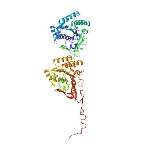
    4IJM

  • PubMed Abstract: 

    The Synechococcus elongatus KaiA, KaiB, and KaiC proteins in the presence of ATP generate a post-translational oscillator that runs in a temperature-compensated manner with a period of 24 h. KaiA dimer stimulates phosphorylation of KaiC hexamer at two sites per subunit, T432 and S431, and KaiB dimers antagonize KaiA action and induce KaiC subunit exchange. Neither the mechanism of KaiA-stimulated KaiC phosphorylation nor that of KaiB-mediated KaiC dephosphorylation is understood in detail at present. We demonstrate here that the A422V KaiC mutant sheds light on the former mechanism. It was previously reported that A422V is less sensitive to dark pulse-induced phase resetting and has a reduced amplitude of the KaiC phosphorylation rhythm in vivo. A422 maps to a loop (422-loop) that continues toward the phosphorylation sites. By pulling on the C-terminal peptide of KaiC (A-loop), KaiA removes restraints from the adjacent 422-loop whose increased flexibility indirectly promotes kinase activity. We found in the crystal structure that A422V KaiC lacks phosphorylation at S431 and exhibits a subtle, local conformational change relative to wild-type KaiC. Molecular dynamics simulations indicate higher mobility of the 422-loop in the absence of the A-loop and mobility differences in other areas associated with phosphorylation activity between wild-type and mutant KaiCs. The A-loop-422-loop relay that informs KaiC phosphorylation sites of KaiA dimer binding propagates to loops from neighboring KaiC subunits, thus providing support for a concerted allosteric mechanism of phosphorylation.


  • Organizational Affiliation

    Department of Biochemistry, Vanderbilt University, School of Medicine, Nashville, TN 37232, USA. martin.egli@vanderbilt.edu


Macromolecules
Find similar proteins by:  (by identity cutoff)  |  3D Structure
Entity ID: 1
MoleculeChains Sequence LengthOrganismDetailsImage
Circadian clock protein kinase KaiC
A, B, C, D, E
A, B, C, D, E, F
512Synechococcus elongatus PCC 7942 = FACHB-805Mutation(s): 1 
Gene Names: kaiCsee0011Synpcc7942_1216
EC: 2.7.11.1
UniProt
Find proteins for Q79PF4 (Synechococcus elongatus (strain ATCC 33912 / PCC 7942 / FACHB-805))
Explore Q79PF4 
Go to UniProtKB:  Q79PF4
Entity Groups  
Sequence Clusters30% Identity50% Identity70% Identity90% Identity95% Identity100% Identity
UniProt GroupQ79PF4
Sequence Annotations
Expand
  • Reference Sequence
Small Molecules
Ligands 3 Unique
IDChains Name / Formula / InChI Key2D Diagram3D Interactions
ATP
Query on ATP

Download Ideal Coordinates CCD File 
AA [auth E]
EA [auth F]
H [auth A]
I [auth A]
J [auth A]
AA [auth E],
EA [auth F],
H [auth A],
I [auth A],
J [auth A],
L [auth B],
M [auth B],
R [auth C],
S [auth C],
V [auth D],
W [auth D],
Z [auth E]
ADENOSINE-5'-TRIPHOSPHATE
C10 H16 N5 O13 P3
ZKHQWZAMYRWXGA-KQYNXXCUSA-N
PO4
Query on PO4

Download Ideal Coordinates CCD File 
BA [auth E]
CA [auth E]
FA [auth F]
GA [auth F]
N [auth B]
BA [auth E],
CA [auth E],
FA [auth F],
GA [auth F],
N [auth B],
O [auth B],
T [auth C],
X [auth D]
PHOSPHATE ION
O4 P
NBIIXXVUZAFLBC-UHFFFAOYSA-K
MG
Query on MG

Download Ideal Coordinates CCD File 
DA [auth F]
G [auth A]
K [auth B]
P [auth B]
Q [auth C]
DA [auth F],
G [auth A],
K [auth B],
P [auth B],
Q [auth C],
U [auth D],
Y [auth E]
MAGNESIUM ION
Mg
JLVVSXFLKOJNIY-UHFFFAOYSA-N
Modified Residues  1 Unique
IDChains TypeFormula2D DiagramParent
TPO
Query on TPO
A, B, C, D, E
A, B, C, D, E, F
L-PEPTIDE LINKINGC4 H10 N O6 PTHR
Experimental Data & Validation

Experimental Data

  • Method: X-RAY DIFFRACTION
  • Resolution: 3.35 Å
  • R-Value Free: 0.261 
  • R-Value Work: 0.217 
  • R-Value Observed: 0.219 
  • Space Group: P 21 21 21
Unit Cell:
Length ( Å )Angle ( ˚ )
a = 131.362α = 90
b = 134.468β = 90
c = 203.936γ = 90
Software Package:
Software NamePurpose
PHENIXrefinement
CNSrefinement
HKL-2000data collection
HKL-2000data reduction
HKL-2000data scaling
CNSphasing

Structure Validation

View Full Validation Report


Ligand Structure Quality Assessment 


Entry History 

Deposition Data

Revision History  (Full details and data files)

  • Version 1.0: 2013-02-13
    Type: Initial release
  • Version 1.1: 2013-03-06
    Changes: Database references
  • Version 1.2: 2019-07-17
    Changes: Data collection, Derived calculations, Refinement description
  • Version 1.3: 2023-09-20
    Changes: Data collection, Database references, Derived calculations, Refinement description