4IGR

Crystal structure of the kainate receptor GluK3 ligand-binding domain in complex with the agonist ZA302


Experimental Data Snapshot

  • Method: X-RAY DIFFRACTION
  • Resolution: 2.65 Å
  • R-Value Free: 0.267 
  • R-Value Work: 0.198 
  • R-Value Observed: 0.201 

wwPDB Validation   3D Report Full Report


Ligand Structure Quality Assessment 


This is version 1.3 of the entry. See complete history


Literature

Chemoenzymatic synthesis of new 2,4-syn-functionalized (S)-glutamate analogues and structure-activity relationship studies at ionotropic glutamate receptors and excitatory amino acid transporters.

Assaf, Z.Larsen, A.P.Venskutonyte, R.Han, L.Abrahamsen, B.Nielsen, B.Gajhede, M.Kastrup, J.S.Jensen, A.A.Pickering, D.S.Frydenvang, K.Gefflaut, T.Bunch, L.

(2013) J Med Chem 56: 1614-1628

  • DOI: https://doi.org/10.1021/jm301433m
  • Primary Citation of Related Structures:  
    4IGR, 4IGT

  • PubMed Abstract: 

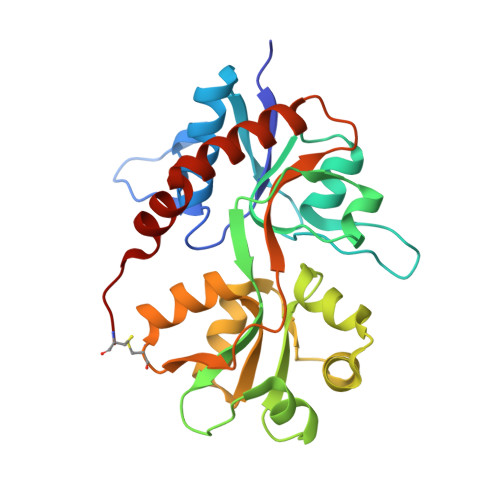
    In the mammalian central nervous system, (S)-glutamate (Glu) is released from the presynaptic neuron where it activates a plethora of pre- and postsynaptic Glu receptors. The fast acting ionotropic Glu receptors (iGluRs) are ligand gated ion channels and are believed to be involved in a vast number of neurological functions such as memory and learning, synaptic plasticity, and motor function. The synthesis of 14 enantiopure 2,4-syn-Glu analogues 2b-p is accessed by a short and efficient chemoenzymatic approach starting from readily available cyclohexanone 3. Pharmacological characterization at the iGluRs and EAAT1-3 subtypes revealed analogue 2i as a selective GluK1 ligand with low nanomolar affinity. Two X-ray crystal structures of the key analogue 2i in the ligand-binding domain (LBD) of GluA2 and GluK3 were determined. Partial domain closure was seen in the GluA2-LBD complex with 2i comparable to that induced by kainate. In contrast, full domain closure was observed in the GluK3-LBD complex with 2i, similar to that of GluK3-LBD with glutamate bound.


  • Organizational Affiliation

    Department of Drug Design and Pharmacology, Faculty of Health and Medical Sciences, University of Copenhagen, 2100 Copenhagen Ø, Denmark.


Macromolecules
Find similar proteins by:  (by identity cutoff)  |  3D Structure
Entity ID: 1
MoleculeChains Sequence LengthOrganismDetailsImage
Glutamate receptor, ionotropic kainate 3258Rattus norvegicusMutation(s): 0 
Gene Names: Glur7Grik3
UniProt
Find proteins for P42264 (Rattus norvegicus)
Explore P42264 
Go to UniProtKB:  P42264
Entity Groups  
Sequence Clusters30% Identity50% Identity70% Identity90% Identity95% Identity100% Identity
UniProt GroupP42264
Sequence Annotations
Expand
  • Reference Sequence
Small Molecules
Binding Affinity Annotations 
IDSourceBinding Affinity
3ZA BindingDB:  4IGR Ki: 123 (nM) from 1 assay(s)
PDBBind:  4IGR Kd: 63 (nM) from 1 assay(s)
Experimental Data & Validation

Experimental Data

  • Method: X-RAY DIFFRACTION
  • Resolution: 2.65 Å
  • R-Value Free: 0.267 
  • R-Value Work: 0.198 
  • R-Value Observed: 0.201 
  • Space Group: P 41 2 2
Unit Cell:
Length ( Å )Angle ( ˚ )
a = 68.18α = 90
b = 68.18β = 90
c = 126.52γ = 90
Software Package:
Software NamePurpose
SCALAdata scaling
PHENIXrefinement
PDB_EXTRACTdata extraction
MOSFLMdata reduction
PHASERphasing

Structure Validation

View Full Validation Report


Ligand Structure Quality Assessment 


Entry History 

Deposition Data

Revision History  (Full details and data files)

  • Version 1.0: 2013-03-06
    Type: Initial release
  • Version 1.1: 2014-09-03
    Changes: Database references
  • Version 1.2: 2017-08-23
    Changes: Data collection, Refinement description, Source and taxonomy
  • Version 1.3: 2023-11-08
    Changes: Data collection, Database references, Derived calculations, Refinement description