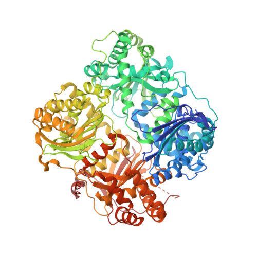
4IFH

Crystal structure of human insulin degrading enzyme (IDE) in complex with compound BDM44619


Experimental Data Snapshot

  • Method: X-RAY DIFFRACTION
  • Resolution: 3.29 Å
  • R-Value Free: 0.224 
  • R-Value Work: 0.182 
  • R-Value Observed: 0.184 

wwPDB Validation   3D Report Full Report


Ligand Structure Quality Assessment 


This is version 1.2 of the entry. See complete history


Literature

Catalytic site inhibition of insulin-degrading enzyme by a small molecule induces glucose intolerance in mice.

Deprez-Poulain, R.Hennuyer, N.Bosc, D.Liang, W.G.Enee, E.Marechal, X.Charton, J.Totobenazara, J.Berte, G.Jahklal, J.Verdelet, T.Dumont, J.Dassonneville, S.Woitrain, E.Gauriot, M.Paquet, C.Duplan, I.Hermant, P.Cantrelle, F.X.Sevin, E.Culot, M.Landry, V.Herledan, A.Piveteau, C.Lippens, G.Leroux, F.Tang, W.J.van Endert, P.Staels, B.Deprez, B.

(2015) Nat Commun 6: 8250-8250

  • DOI: https://doi.org/10.1038/ncomms9250
  • Primary Citation of Related Structures:  
    4IFH, 4NXO, 4RE9

  • PubMed Abstract: 

    Insulin-degrading enzyme (IDE) is a protease that cleaves insulin and other bioactive peptides such as amyloid-β. Knockout and genetic studies have linked IDE to Alzheimer's disease and type-2 diabetes. As the major insulin-degrading protease, IDE is a candidate drug target in diabetes. Here we have used kinetic target-guided synthesis to design the first catalytic site inhibitor of IDE suitable for in vivo studies (BDM44768). Crystallographic and small angle X-ray scattering analyses show that it locks IDE in a closed conformation. Among a panel of metalloproteases, BDM44768 selectively inhibits IDE. Acute treatment of mice with BDM44768 increases insulin signalling and surprisingly impairs glucose tolerance in an IDE-dependent manner. These results confirm that IDE is involved in pathways that modulate short-term glucose homeostasis, but casts doubt on the general usefulness of the inhibition of IDE catalytic activity to treat diabetes.


  • Organizational Affiliation

    Institut Pasteur de Lille, Lille F-59000, France.


Macromolecules
Find similar proteins by:  (by identity cutoff)  |  3D Structure
Entity ID: 1
MoleculeChains Sequence LengthOrganismDetailsImage
Insulin-degrading enzyme
A, B
990Homo sapiensMutation(s): 13 
Gene Names: IDE
EC: 3.4.24.56
UniProt & NIH Common Fund Data Resources
Find proteins for P14735 (Homo sapiens)
Explore P14735 
Go to UniProtKB:  P14735
PHAROS:  P14735
GTEx:  ENSG00000119912 
Entity Groups  
Sequence Clusters30% Identity50% Identity70% Identity90% Identity95% Identity100% Identity
UniProt GroupP14735
Sequence Annotations
Expand
  • Reference Sequence
Small Molecules
Ligands 2 Unique
IDChains Name / Formula / InChI Key2D Diagram3D Interactions
1EF
Query on 1EF

Download Ideal Coordinates CCD File 
E [auth B]N-({1-[(2R)-4-(hydroxyamino)-1-(naphthalen-2-yl)-4-oxobutan-2-yl]-1H-1,2,3-triazol-4-yl}methyl)-4-methylbenzamide
C25 H25 N5 O3
QIIHKUBWBJHTFJ-HSZRJFAPSA-N
ZN
Query on ZN

Download Ideal Coordinates CCD File 
C [auth A],
D [auth B]
ZINC ION
Zn
PTFCDOFLOPIGGS-UHFFFAOYSA-N
Experimental Data & Validation

Experimental Data

  • Method: X-RAY DIFFRACTION
  • Resolution: 3.29 Å
  • R-Value Free: 0.224 
  • R-Value Work: 0.182 
  • R-Value Observed: 0.184 
  • Space Group: P 65
Unit Cell:
Length ( Å )Angle ( ˚ )
a = 263.022α = 90
b = 263.022β = 90
c = 90.59γ = 120
Software Package:
Software NamePurpose
HKL-3000data collection
PHASERphasing
PHENIXrefinement
HKL-3000data reduction
HKL-3000data scaling

Structure Validation

View Full Validation Report


Ligand Structure Quality Assessment 


Entry History 

Deposition Data

Revision History  (Full details and data files)

  • Version 1.0: 2013-12-18
    Type: Initial release
  • Version 1.1: 2015-10-28
    Changes: Database references
  • Version 1.2: 2024-02-28
    Changes: Data collection, Database references, Derived calculations