4I8O

Crystal structure of the toxin RnlA from Escherichia coli


Experimental Data Snapshot

  • Method: X-RAY DIFFRACTION
  • Resolution: 2.10 Å
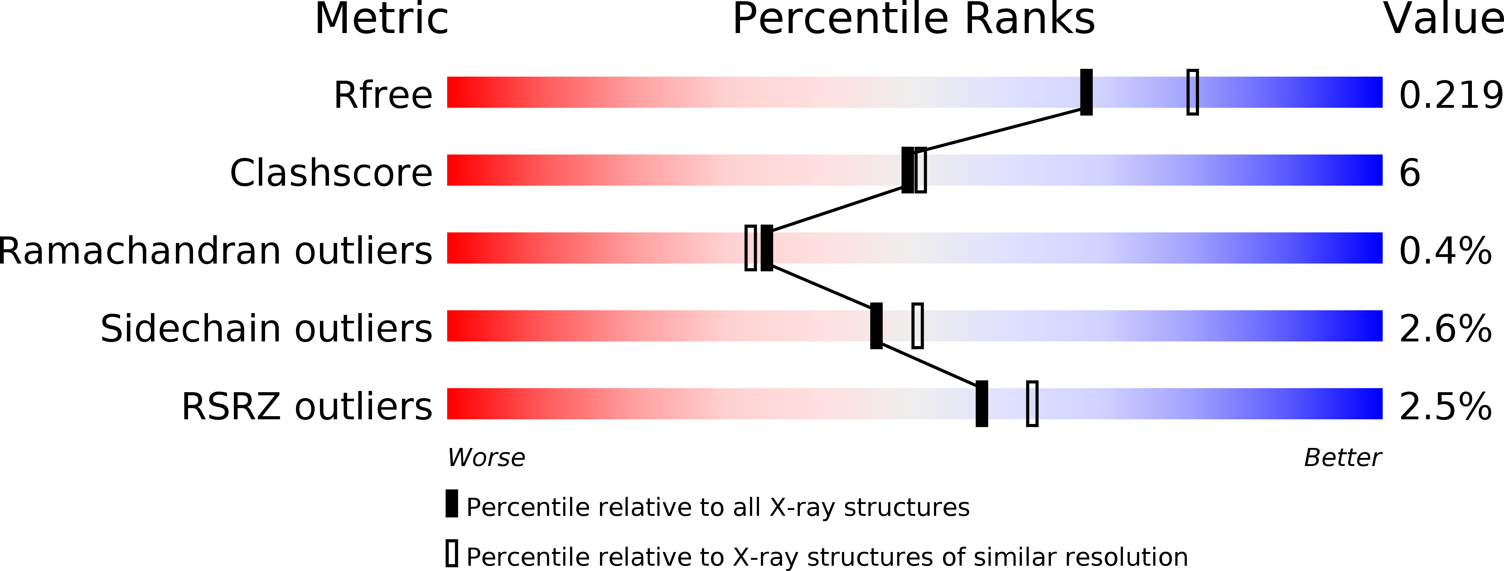
  • R-Value Free: 0.229 
  • R-Value Work: 0.196 
  • R-Value Observed: 0.196 

wwPDB Validation   3D Report Full Report


This is version 1.3 of the entry. See complete history


Literature

Structure-function studies of Escherichia coli RnlA reveal a novel toxin structure involved in bacteriophage resistance.

Wei, Y.Gao, Z.Q.Otsuka, Y.Naka, K.Yonesaki, T.Zhang, H.Dong, Y.H.

(2013) Mol Microbiol 90: 956-965

  • DOI: https://doi.org/10.1111/mmi.12409
  • Primary Citation of Related Structures:  
    4I8O

  • PubMed Abstract: 

    Escherichia coli RnlA-RnlB is a newly identified toxin-antitoxin (TA) system that plays a role in bacteriophage resistance. RnlA functions as a toxin with mRNA endoribonuclease activity and the cognate antitoxin RnlB inhibits RnlA toxicity in E. coli cells. Interestingly, T4 phage encodes the antitoxin Dmd, which acts against RnlA to promote its own propagation, suggesting that RnlA-Dmd represents a novel TA system. Here, we have determined the crystal structure of RnlA refined to 2.10  (Dmd-binding domain), which is an organization not previously observed among known toxin structures. Small-angle X-ray scattering (SAXS) analysis revealed that RnlA forms a dimer in solution via interactions between the DBDs from both monomers. The in vitro and in vivo functional studies showed that among the three domains, only the DBD is responsible for recognition and inhibition by Dmd and subcellular location of RnlA. In particular, the helix located at the C-terminus of DBD plays a vital role in binding Dmd. Our comprehensive studies reveal the key region responsible for RnlA toxicity and provide novel insights into its structure-function relationship.


  • Organizational Affiliation

    School of Life Sciences, University of Science and Technology of China, Hefei, 230027, China; Beijing Synchrotron Radiation Facility, Institute of High Energy Physics, Chinese Academy of Sciences, Beijing, 100049, China.


Macromolecules
Find similar proteins by:  (by identity cutoff)  |  3D Structure
Entity ID: 1
MoleculeChains Sequence LengthOrganismDetailsImage
TOXIN RNLA
A, B
358Escherichia coli K-12Mutation(s): 0 
Gene Names: yfjNb2630JW2611
UniProt
Find proteins for P52129 (Escherichia coli (strain K12))
Explore P52129 
Go to UniProtKB:  P52129
Entity Groups  
Sequence Clusters30% Identity50% Identity70% Identity90% Identity95% Identity100% Identity
UniProt GroupP52129
Sequence Annotations
Expand
  • Reference Sequence
Small Molecules
Modified Residues  1 Unique
IDChains TypeFormula2D DiagramParent
MSE
Query on MSE
A, B
L-PEPTIDE LINKINGC5 H11 N O2 SeMET
Experimental Data & Validation

Experimental Data

  • Method: X-RAY DIFFRACTION
  • Resolution: 2.10 Å
  • R-Value Free: 0.229 
  • R-Value Work: 0.196 
  • R-Value Observed: 0.196 
  • Space Group: P 1
Unit Cell:
Length ( Å )Angle ( ˚ )
a = 57.207α = 69.24
b = 64.321β = 79.53
c = 82.623γ = 75.03
Software Package:
Software NamePurpose
MAR345data collection
SOLVEphasing
PHENIXrefinement
ADSCdata collection
HKL-2000data reduction
SCALEPACKdata scaling

Structure Validation

View Full Validation Report


Entry History 

Deposition Data

Revision History  (Full details and data files)

  • Version 1.0: 2013-10-23
    Type: Initial release
  • Version 1.1: 2013-12-11
    Changes: Database references
  • Version 1.2: 2014-12-03
    Changes: Structure summary
  • Version 1.3: 2017-11-15
    Changes: Refinement description