


X-ray structure of the unliganded uridine phosphorylase from Yersinia pseudotuberculosis at 2.12A resolution
Lashkov, A.A., Balaev, V.V., Prokofev, I.I., Betzel, C., Gabdoulkhakov, A.G., Mikhailov, A.M.To be published.
Experimental Data Snapshot
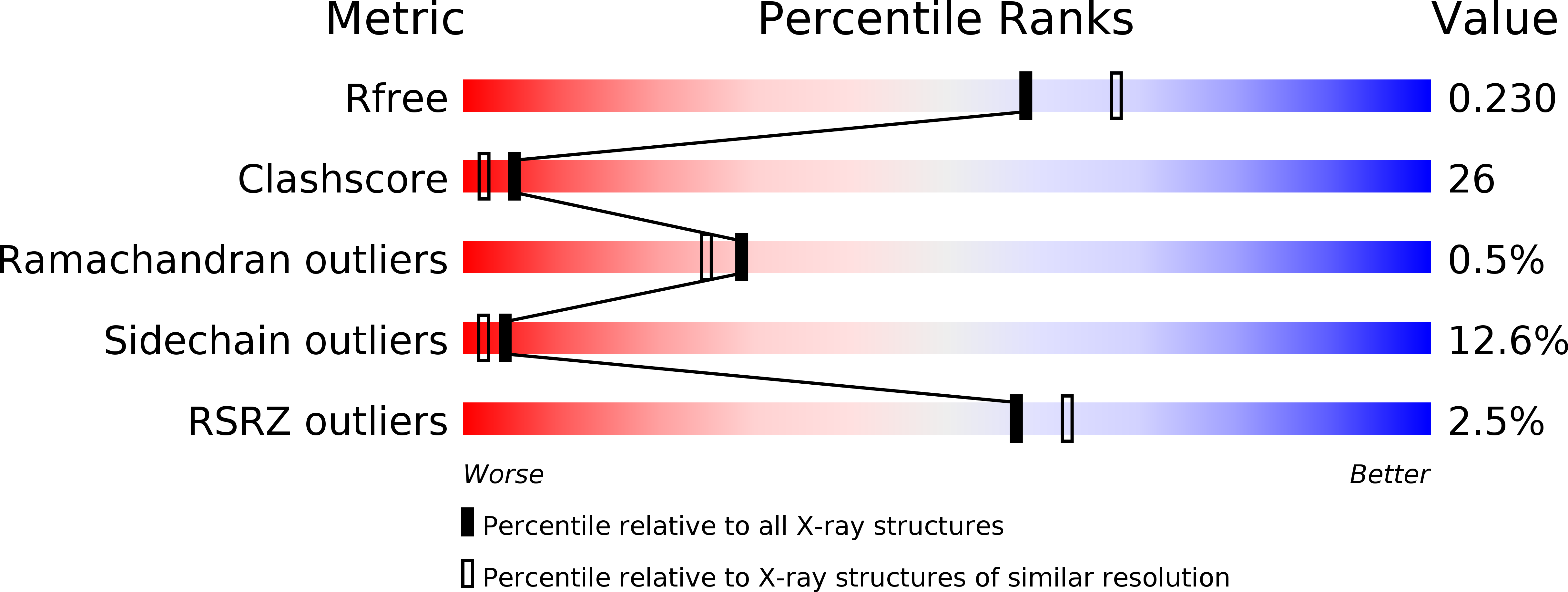
wwPDB Validation 3D Report Full Report
Entity ID: 1 | |||||
|---|---|---|---|---|---|
| Molecule | Chains | Sequence Length | Organism | Details | Image |
| Uridine phosphorylase | 253 | Yersinia pseudotuberculosis | Mutation(s): 0 Gene Names: udp EC: 2.4.2.3 | 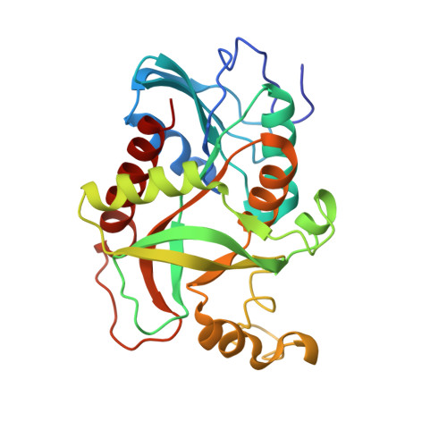 | |
UniProt | |||||
Find proteins for Q7AZX0 (Yersinia pseudotuberculosis) Explore Q7AZX0 Go to UniProtKB: Q7AZX0 | |||||
Entity Groups | |||||
| Sequence Clusters | 30% Identity50% Identity70% Identity90% Identity95% Identity100% Identity | ||||
| UniProt Group | Q7AZX0 | ||||
Sequence AnnotationsExpand | |||||
| |||||
| Length ( Å ) | Angle ( ˚ ) |
|---|---|
| a = 156.68 | α = 90 |
| b = 156.68 | β = 90 |
| c = 48.46 | γ = 120 |
| Software Name | Purpose |
|---|---|
| SCALA | data scaling |
| PHASER | phasing |
| PHENIX | refinement |
| PDB_EXTRACT | data extraction |
| HKL-2000 | data collection |
| XSCALE | data scaling |
RCSB PDB (citation) is hosted by
RCSB PDB is a member of the

