4HI0

Crystal Structure of Helicobacter pylori Urease Accessory Protein UreF/H/G complex


Experimental Data Snapshot

  • Method: X-RAY DIFFRACTION
  • Resolution: 2.35 Å
  • R-Value Free: 0.242 
  • R-Value Work: 0.192 
  • R-Value Observed: 0.193 

wwPDB Validation   3D Report Full Report


Ligand Structure Quality Assessment 


This is version 1.2 of the entry. See complete history


Literature

Structure of UreG/UreF/UreH complex reveals how urease accessory proteins facilitate maturation of Helicobacter pylori urease.

Fong, Y.H.Wong, H.C.Yuen, M.H.Lau, P.H.Chen, Y.W.Wong, K.B.

(2013) PLoS Biol 11: e1001678-e1001678

  • DOI: https://doi.org/10.1371/journal.pbio.1001678
  • Primary Citation of Related Structures:  
    4HI0

  • PubMed Abstract: 

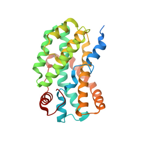
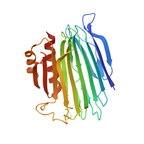
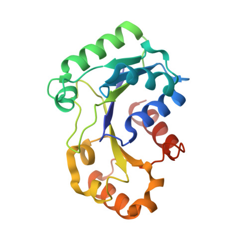
    Urease is a metalloenzyme essential for the survival of Helicobacter pylori in acidic gastric environment. Maturation of urease involves carbamylation of Lys219 and insertion of two nickel ions at its active site. This process requires GTP hydrolysis and the formation of a preactivation complex consisting of apo-urease and urease accessory proteins UreF, UreH, and UreG. UreF and UreH form a complex to recruit UreG, which is a SIMIBI class GTPase, to the preactivation complex. We report here the crystal structure of the UreG/UreF/UreH complex, which illustrates how UreF and UreH facilitate dimerization of UreG, and assembles its metal binding site by juxtaposing two invariant Cys66-Pro67-His68 metal binding motif at the interface to form the (UreG/UreF/UreH)2 complex. Interaction studies revealed that addition of nickel and GTP to the UreG/UreF/UreH complex releases a UreG dimer that binds a nickel ion at the dimeric interface. Substitution of Cys66 and His68 with alanine abolishes the formation of the nickel-charged UreG dimer. This nickel-charged UreG dimer can activate urease in vitro in the presence of the UreF/UreH complex. Static light scattering and atomic absorption spectroscopy measurements demonstrated that the nickel-charged UreG dimer, upon GTP hydrolysis, reverts to its monomeric form and releases nickel to urease. Based on our results, we propose a mechanism on how urease accessory proteins facilitate maturation of urease.


  • Organizational Affiliation

    School of Life Sciences, Center for Protein Science and Crystallography, The Chinese University of Hong Kong, Hong Kong, China.


Macromolecules
Find similar proteins by:  (by identity cutoff)  |  3D Structure
Entity ID: 1
MoleculeChains Sequence LengthOrganismDetailsImage
Urease accessory protein UreF
A, C
254Helicobacter pylori 26695Mutation(s): 0 
Gene Names: HP_0069ureF
UniProt
Find proteins for Q09065 (Helicobacter pylori (strain ATCC 700392 / 26695))
Explore Q09065 
Go to UniProtKB:  Q09065
Entity Groups  
Sequence Clusters30% Identity50% Identity70% Identity90% Identity95% Identity100% Identity
UniProt GroupQ09065
Sequence Annotations
Expand
  • Reference Sequence
Find similar proteins by:  (by identity cutoff)  |  3D Structure
Entity ID: 2
MoleculeChains Sequence LengthOrganismDetailsImage
Urease accessory protein UreH
B, D
265Helicobacter pylori 26695Mutation(s): 0 
Gene Names: HP_0067ureH
UniProt
Find proteins for Q09067 (Helicobacter pylori (strain ATCC 700392 / 26695))
Explore Q09067 
Go to UniProtKB:  Q09067
Entity Groups  
Sequence Clusters30% Identity50% Identity70% Identity90% Identity95% Identity100% Identity
UniProt GroupQ09067
Sequence Annotations
Expand
  • Reference Sequence
Find similar proteins by:  (by identity cutoff)  |  3D Structure
Entity ID: 3
MoleculeChains Sequence LengthOrganismDetailsImage
Urease accessory protein UreG
E, F
199Helicobacter pylori 26695Mutation(s): 0 
Gene Names: HP_0068ureG
UniProt
Find proteins for Q09066 (Helicobacter pylori (strain ATCC 700392 / 26695))
Explore Q09066 
Go to UniProtKB:  Q09066
Entity Groups  
Sequence Clusters30% Identity50% Identity70% Identity90% Identity95% Identity100% Identity
UniProt GroupQ09066
Sequence Annotations
Expand
  • Reference Sequence
Small Molecules
Experimental Data & Validation

Experimental Data

  • Method: X-RAY DIFFRACTION
  • Resolution: 2.35 Å
  • R-Value Free: 0.242 
  • R-Value Work: 0.192 
  • R-Value Observed: 0.193 
  • Space Group: P 21 21 21
Unit Cell:
Length ( Å )Angle ( ˚ )
a = 76.124α = 90
b = 96.429β = 90
c = 237.452γ = 90
Software Package:
Software NamePurpose
GDAdata collection
PHASERphasing
PHENIXrefinement
XDSdata reduction
XDSdata scaling

Structure Validation

View Full Validation Report


Ligand Structure Quality Assessment 


Entry History 

Deposition Data

Revision History  (Full details and data files)

  • Version 1.0: 2013-10-16
    Type: Initial release
  • Version 1.1: 2014-01-15
    Changes: Database references
  • Version 1.2: 2024-02-28
    Changes: Data collection, Database references, Derived calculations