4HH1

Dark-state structure of AppA wild-type without the Cys-rich region from Rb. sphaeroides


Experimental Data Snapshot

  • Method: X-RAY DIFFRACTION
  • Resolution: 3.50 Å
  • R-Value Free: 0.226 
  • R-Value Work: 0.186 
  • R-Value Observed: 0.188 

wwPDB Validation   3D Report Full Report


Ligand Structure Quality Assessment 


This is version 1.4 of the entry. See complete history


Literature

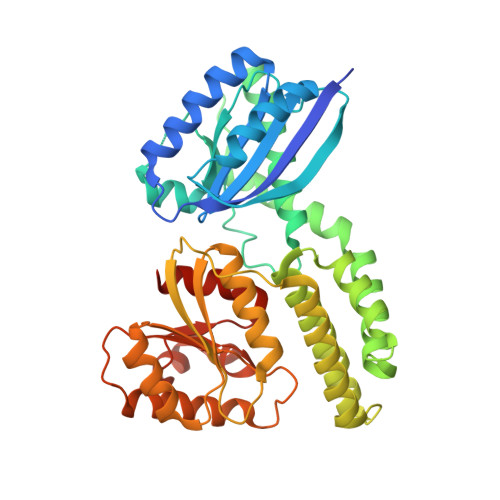
A ternary AppA-PpsR-DNA complex mediates light regulation of photosynthesis-related gene expression.

Winkler, A.Heintz, U.Lindner, R.Reinstein, J.Shoeman, R.L.Schlichting, I.

(2013) Nat Struct Mol Biol 20: 859-867

  • DOI: https://doi.org/10.1038/nsmb.2597
  • Primary Citation of Related Structures:  
    4HH0, 4HH1, 4HH2, 4HH3

  • PubMed Abstract: 

    The anoxygenic phototrophic bacterium Rhodobacter sphaeroides uses different energy sources, depending on environmental conditions including aerobic respiration or, in the absence of oxygen, photosynthesis. Photosynthetic genes are repressed at high oxygen tension, but at intermediate levels their partial expression prepares the bacterium for using light energy. Illumination, however, enhances repression under semiaerobic conditions. Here, we describe molecular details of two proteins mediating oxygen and light control of photosynthesis-gene expression: the light-sensing antirepressor AppA and the transcriptional repressor PpsR. Our crystal structures of both proteins and their complex and hydrogen/deuterium-exchange data show that light activation of AppA-PpsR2 affects the PpsR effector region within the complex. DNA binding studies demonstrate the formation of a light-sensitive ternary AppA-PpsR-DNA complex. We discuss implications of these results for regulation by light and oxygen, highlighting new insights into blue light-mediated signal transduction.


  • Organizational Affiliation

    Department of Biomolecular Mechanisms, Max Planck Institute for Medical Research, Heidelberg, Germany. andreas.winkler@mpimf-heidelberg.mpg.de


Macromolecules
Find similar proteins by:  (by identity cutoff)  |  3D Structure
Entity ID: 1
MoleculeChains Sequence LengthOrganismDetailsImage
AppA protein
A, B
406Cereibacter sphaeroidesMutation(s): 1 
Gene Names: appA
UniProt
Find proteins for Q53119 (Cereibacter sphaeroides)
Explore Q53119 
Go to UniProtKB:  Q53119
Entity Groups  
Sequence Clusters30% Identity50% Identity70% Identity90% Identity95% Identity100% Identity
UniProt GroupQ53119
Sequence Annotations
Expand
  • Reference Sequence
Small Molecules
Experimental Data & Validation

Experimental Data

  • Method: X-RAY DIFFRACTION
  • Resolution: 3.50 Å
  • R-Value Free: 0.226 
  • R-Value Work: 0.186 
  • R-Value Observed: 0.188 
  • Space Group: P 32 1 2
Unit Cell:
Length ( Å )Angle ( ˚ )
a = 70.53α = 90
b = 70.53β = 90
c = 383.81γ = 120
Software Package:
Software NamePurpose
PHENIXmodel building
PHENIXrefinement
XDSdata reduction
XSCALEdata scaling
PHENIXphasing

Structure Validation

View Full Validation Report


Ligand Structure Quality Assessment 


Entry History 

Deposition Data

Revision History  (Full details and data files)

  • Version 1.0: 2013-06-05
    Type: Initial release
  • Version 1.1: 2013-07-10
    Changes: Database references
  • Version 1.2: 2013-07-17
    Changes: Database references
  • Version 1.3: 2017-11-15
    Changes: Refinement description
  • Version 1.4: 2024-02-28
    Changes: Data collection, Database references, Derived calculations