4H7U

Crystal structure of pyranose dehydrogenase from Agaricus meleagris, wildtype


Experimental Data Snapshot

  • Method: X-RAY DIFFRACTION
  • Resolution: 1.60 Å
  • R-Value Free: 0.201 
  • R-Value Work: 0.171 
  • R-Value Observed: 0.172 

wwPDB Validation   3D Report Full Report


Ligand Structure Quality Assessment 


This is version 2.1 of the entry. See complete history


Literature

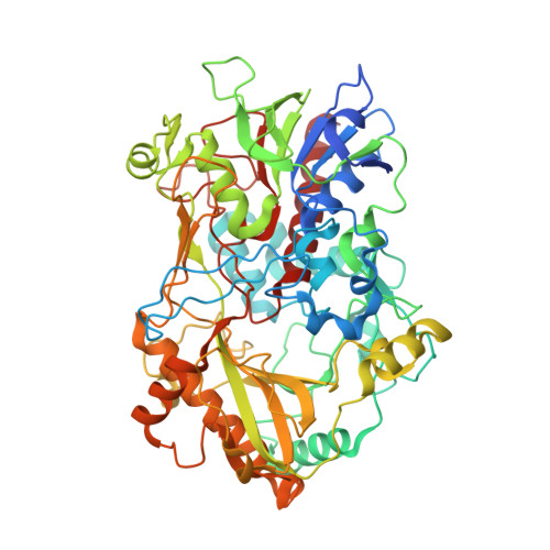
The 1.6 A crystal structure of pyranose dehydrogenase from Agaricus meleagris rationalizes substrate specificity and reveals a flavin intermediate.

Tan, T.C.Spadiut, O.Wongnate, T.Sucharitakul, J.Krondorfer, I.Sygmund, C.Haltrich, D.Chaiyen, P.Peterbauer, C.K.Divne, C.

(2013) PLoS One 8: e53567-e53567

  • DOI: https://doi.org/10.1371/journal.pone.0053567
  • Primary Citation of Related Structures:  
    4H7U

  • PubMed Abstract: 

    Pyranose dehydrogenases (PDHs) are extracellular flavin-dependent oxidoreductases secreted by litter-decomposing fungi with a role in natural recycling of plant matter. All major monosaccharides in lignocellulose are oxidized by PDH at comparable yields and efficiencies. Oxidation takes place as single-oxidation or sequential double-oxidation reactions of the carbohydrates, resulting in sugar derivatives oxidized primarily at C2, C3 or C2/3 with the concomitant reduction of the flavin. A suitable electron acceptor then reoxidizes the reduced flavin. Whereas oxygen is a poor electron acceptor for PDH, several alternative acceptors, e.g., quinone compounds, naturally present during lignocellulose degradation, can be used. We have determined the 1.6-Å crystal structure of PDH from Agaricus meleagris. Interestingly, the flavin ring in PDH is modified by a covalent mono- or di-atomic species at the C(4a) position. Under normal conditions, PDH is not oxidized by oxygen; however, the related enzyme pyranose 2-oxidase (P2O) activates oxygen by a mechanism that proceeds via a covalent flavin C(4a)-hydroperoxide intermediate. Although the flavin C(4a) adduct is common in monooxygenases, it is unusual for flavoprotein oxidases, and it has been proposed that formation of the intermediate would be unfavorable in these oxidases. Thus, the flavin adduct in PDH not only shows that the adduct can be favorably accommodated in the active site, but also provides important details regarding the structural, spatial and physicochemical requirements for formation of this flavin intermediate in related oxidases. Extensive in silico modeling of carbohydrates in the PDH active site allowed us to rationalize the previously reported patterns of substrate specificity and regioselectivity. To evaluate the regioselectivity of D-glucose oxidation, reduction experiments were performed using fluorinated glucose. PDH was rapidly reduced by 3-fluorinated glucose, which has the C2 position accessible for oxidation, whereas 2-fluorinated glucose performed poorly (C3 accessible), indicating that the glucose C2 position is the primary site of attack.


  • Organizational Affiliation

    School of Biotechnology, KTH Royal Institute of Technology, Stockholm, Sweden.


Macromolecules
Find similar proteins by:  (by identity cutoff)  |  3D Structure
Entity ID: 1
MoleculeChains Sequence LengthOrganismDetailsImage
Pyranose dehydrogenase602Leucoagaricus meleagrisMutation(s): 0 
Gene Names: pdh1
EC: 1.1.99.29
UniProt
Find proteins for Q3L245 (Leucoagaricus meleagris)
Explore Q3L245 
Go to UniProtKB:  Q3L245
Entity Groups  
Sequence Clusters30% Identity50% Identity70% Identity90% Identity95% Identity100% Identity
UniProt GroupQ3L245
Sequence Annotations
Expand
  • Reference Sequence
Oligosaccharides

Help

Entity ID: 2
MoleculeChains Length2D Diagram Glycosylation3D Interactions
2-acetamido-2-deoxy-beta-D-glucopyranose-(1-4)-2-acetamido-2-deoxy-beta-D-glucopyranose
B
2N-Glycosylation
Glycosylation Resources
GlyTouCan:  G42666HT
GlyCosmos:  G42666HT
GlyGen:  G42666HT
Small Molecules
Ligands 3 Unique
IDChains Name / Formula / InChI Key2D Diagram3D Interactions
FED
Query on FED

Download Ideal Coordinates CCD File 
C [auth A][(2R,3S,4R,5R)-5-(6-amino-9H-purin-9-yl)-3,4-dihydroxytetrahydrofuran-2-yl]methyl (2R,3S,4S)-2,3,4-trihydroxy-5-[(4aR)-4a-hydroxy-7,8-dimethyl-2,4-dioxo-3,4,4a,5-tetrahydrobenzo[g]pteridin-10(2H)-yl]pentyl dihydrogen diphosphate (non-preferred name)
C27 H35 N9 O16 P2
WTOYVZYXGAOYIM-QUWWARRSSA-N
NAG
Query on NAG

Download Ideal Coordinates CCD File 
D [auth A]2-acetamido-2-deoxy-beta-D-glucopyranose
C8 H15 N O6
OVRNDRQMDRJTHS-FMDGEEDCSA-N
PO4
Query on PO4

Download Ideal Coordinates CCD File 
E [auth A]PHOSPHATE ION
O4 P
NBIIXXVUZAFLBC-UHFFFAOYSA-K
Experimental Data & Validation

Experimental Data

  • Method: X-RAY DIFFRACTION
  • Resolution: 1.60 Å
  • R-Value Free: 0.201 
  • R-Value Work: 0.171 
  • R-Value Observed: 0.172 
  • Space Group: P 21 21 21
Unit Cell:
Length ( Å )Angle ( ˚ )
a = 51.938α = 90
b = 74.64β = 90
c = 139.286γ = 90
Software Package:
Software NamePurpose
MAR345data collection
BALBESphasing
REFMACrefinement
XDSdata reduction
XSCALEdata scaling

Structure Validation

View Full Validation Report


Ligand Structure Quality Assessment 


Entry History 

Deposition Data

Revision History  (Full details and data files)

  • Version 1.0: 2013-08-07
    Type: Initial release
  • Version 1.1: 2017-11-15
    Changes: Data collection, Refinement description
  • Version 2.0: 2020-07-29
    Type: Remediation
    Reason: Carbohydrate remediation
    Changes: Atomic model, Data collection, Derived calculations, Structure summary
  • Version 2.1: 2023-09-20
    Changes: Data collection, Database references, Refinement description, Structure summary