4GYZ

Mus Musculus Tdp2 Bound to dAMP and Mg2+


Experimental Data Snapshot

  • Method: X-RAY DIFFRACTION
  • Resolution: 2.56 Å
  • R-Value Free: 0.220 
  • R-Value Work: 0.191 
  • R-Value Observed: 0.193 

wwPDB Validation   3D Report Full Report


Ligand Structure Quality Assessment 


This is version 1.5 of the entry. See complete history


Literature

Mechanism of repair of 5'-topoisomerase II-DNA adducts by mammalian tyrosyl-DNA phosphodiesterase 2.

Schellenberg, M.J.Appel, C.D.Adhikari, S.Robertson, P.D.Ramsden, D.A.Williams, R.S.

(2012) Nat Struct Mol Biol 19: 1363-1371

  • DOI: https://doi.org/10.1038/nsmb.2418
  • Primary Citation of Related Structures:  
    4GYZ, 4GZ0, 4GZ1, 4GZ2

  • PubMed Abstract: 

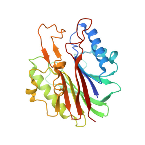
    The topoisomerase II (topo II) DNA incision-and-ligation cycle can be poisoned (for example following treatment with cancer chemotherapeutics) to generate cytotoxic DNA double-strand breaks (DSBs) with topo II covalently conjugated to DNA. Tyrosyl-DNA phosphodiesterase 2 (Tdp2) protects genomic integrity by reversing 5'-phosphotyrosyl-linked topo II-DNA adducts. Here, X-ray structures of mouse Tdp2-DNA complexes reveal that Tdp2 β-2-helix-β DNA damage-binding 'grasp', helical 'cap' and DNA lesion-binding elements fuse to form an elongated protein-DNA conjugate substrate-interaction groove. The Tdp2 DNA-binding surface is highly tailored for engagement of 5'-adducted single-stranded DNA ends and restricts nonspecific endonucleolytic or exonucleolytic processing. Structural, mutational and functional analyses support a single-metal ion catalytic mechanism for the exonuclease-endonuclease-phosphatase (EEP) nuclease superfamily and establish a molecular framework for targeted small-molecule blockade of Tdp2-mediated resistance to anticancer topoisomerase drugs.


  • Organizational Affiliation

    Laboratory of Structural Biology, National Institute of Environmental Health Sciences, US National Institutes of Health, Department of Health and Human Services, North Carolina, USA.


Macromolecules
Find similar proteins by:  (by identity cutoff)  |  3D Structure
Entity ID: 1
MoleculeChains Sequence LengthOrganismDetailsImage
Tyrosyl-DNA phosphodiesterase 2
A, B, C, D, E
A, B, C, D, E, F, G, H, I
256Mus musculusMutation(s): 0 
Gene Names: Tdp2Ttrap
EC: 3.1.4
UniProt
Find proteins for Q9JJX7 (Mus musculus)
Explore Q9JJX7 
Go to UniProtKB:  Q9JJX7
Entity Groups  
Sequence Clusters30% Identity50% Identity70% Identity90% Identity95% Identity100% Identity
UniProt GroupQ9JJX7
Sequence Annotations
Expand
  • Reference Sequence
Small Molecules
Ligands 4 Unique
IDChains Name / Formula / InChI Key2D Diagram3D Interactions
D5M
Query on D5M

Download Ideal Coordinates CCD File 
J [auth A]
L [auth B]
O [auth C]
R [auth D]
T [auth E]
J [auth A],
L [auth B],
O [auth C],
R [auth D],
T [auth E],
Y [auth H]
2'-DEOXYADENOSINE-5'-MONOPHOSPHATE
C10 H14 N5 O6 P
KHWCHTKSEGGWEX-RRKCRQDMSA-N
PO4
Query on PO4

Download Ideal Coordinates CCD File 
V [auth F]PHOSPHATE ION
O4 P
NBIIXXVUZAFLBC-UHFFFAOYSA-K
CL
Query on CL

Download Ideal Coordinates CCD File 
AA [auth I],
N [auth C],
Q [auth D]
CHLORIDE ION
Cl
VEXZGXHMUGYJMC-UHFFFAOYSA-M
MG
Query on MG

Download Ideal Coordinates CCD File 
BA [auth I]
K [auth A]
M [auth B]
P [auth C]
S [auth D]
BA [auth I],
K [auth A],
M [auth B],
P [auth C],
S [auth D],
U [auth E],
W [auth F],
X [auth G],
Z [auth H]
MAGNESIUM ION
Mg
JLVVSXFLKOJNIY-UHFFFAOYSA-N
Experimental Data & Validation

Experimental Data

  • Method: X-RAY DIFFRACTION
  • Resolution: 2.56 Å
  • R-Value Free: 0.220 
  • R-Value Work: 0.191 
  • R-Value Observed: 0.193 
  • Space Group: P 21 21 21
Unit Cell:
Length ( Å )Angle ( ˚ )
a = 85.347α = 90
b = 169.326β = 90
c = 184.959γ = 90
Software Package:
Software NamePurpose
PHENIXrefinement
REFMACrefinement
PDB_EXTRACTdata extraction
HKL-2000data collection
DENZOdata reduction
REFMACphasing
SCALEPACKdata scaling

Structure Validation

View Full Validation Report


Ligand Structure Quality Assessment 


Entry History 

Deposition Data

Revision History  (Full details and data files)

  • Version 1.0: 2012-10-31
    Type: Initial release
  • Version 1.1: 2012-11-21
    Changes: Database references
  • Version 1.2: 2012-11-28
    Changes: Database references
  • Version 1.3: 2013-01-02
    Changes: Database references
  • Version 1.4: 2019-07-17
    Changes: Data collection, Refinement description
  • Version 1.5: 2024-02-28
    Changes: Data collection, Database references, Derived calculations