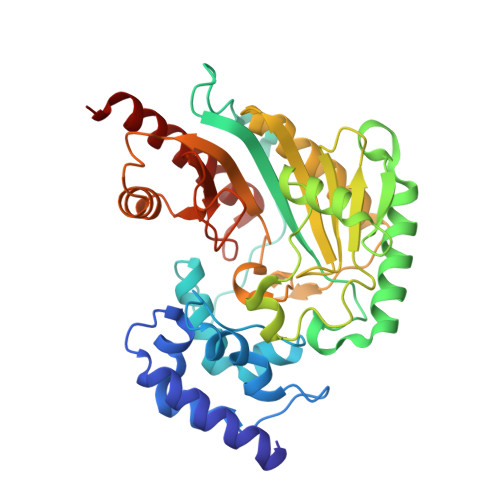
Crystal structures of arginine kinase in complex with ADP, nitrate, and various phosphagen analogs.
Clark, S.A., Davulcu, O., Chapman, M.S.(2012) Biochem Biophys Res Commun 427: 212-217
- PubMed: 22995310
- DOI: https://doi.org/10.1016/j.bbrc.2012.09.053
- Primary Citation of Related Structures:
4GVY, 4GVZ, 4GW0, 4GW2 - PubMed Abstract:
Arginine kinase catalyzes the reversible transfer of a phosphoryl group between ATP and l-arginine and is a monomeric homolog of the human enzyme creatine kinase. Arginine and creatine kinases belongs to the phosphagen kinase family of enzymes, which consists of eight known members, each of which is specific for its own phosphagen. Here, the source of phosphagen specificity in arginine kinase is investigated through the use of phosphagen analogs. Crystal structures have been determined for Limulus polyphemus arginine kinase with one of four arginine analogs bound in a transition state analog complex: l-ornithine, l-citrulline, imino-l-ornithine, and d-arginine. In all complexes, the enzyme achieves a closed conformation very similar to that of the cognate transition state analog complex, but differences are observed in the configurations of bound ligands. Arginine kinase exhibits no detectable activity towards ornithine, citrulline, or imino-l-ornithine, and only trace activity towards d-arginine. The crystal structures presented here demonstrate that phosphagen specificity is derived neither from a lock-and-key mechanism nor a modulation of induced-fit conformational changes, but potentially from subtle distortions in bound substrate configurations.
Organizational Affiliation:
Institute of Molecular Biophysics, Department of Chemistry & Biochemistry, Florida State University, Tallahassee, FL 32306-4380, USA.