4GIG

crystal structure of T69A mutant of trapped Dnae intein precursor


Experimental Data Snapshot

  • Method: X-RAY DIFFRACTION
  • Resolution: 1.80 Å
  • R-Value Free: 0.268 
  • R-Value Work: 0.226 
  • R-Value Observed: 0.226 

wwPDB Validation   3D Report Full Report


This is version 1.2 of the entry. See complete history


Literature

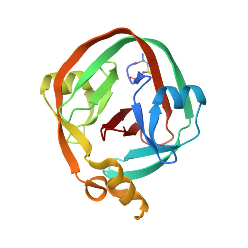
A conserved threonine spring-loads precursor for intein splicing.

Dearden, A.K.Callahan, B.Roey, P.V.Li, Z.Kumar, U.Belfort, M.Nayak, S.K.

(2013) Protein Sci 22: 557-563

  • DOI: https://doi.org/10.1002/pro.2236
  • Primary Citation of Related Structures:  
    4GIG

  • PubMed Abstract: 

    Protein splicing is an autocatalytic process where an "intein" self-cleaves from a precursor and ligates the flanking N- and C-"extein" polypeptides. Inteins occur in all domains of life and have myriad uses in biotechnology. Although the reaction steps of protein splicing are known, mechanistic details remain incomplete, particularly the initial peptide rearrangement at the N-terminal extein/intein junction. Recently, we proposed that this transformation, an N-S acyl shift, is accelerated by a localized conformational strain, between the intein's catalytic cysteine (Cys1) and the neighboring glycine (Gly-1) in the N-extein. That proposal was based on the crystal structure of a catalytically competent trapped precursor. Here, we define the structural origins and mechanistic relevance of the conformational strain using a combination of quantum mechanical simulations, mutational analysis, and X-ray crystallography. Our results implicate a conserved, but largely unstudied, threonine residue of the Ssp DnaE intein (Thr69) as the mediator of conformational strain through hydrogen bonding. Further, the strain imposed by this residue is shown to position the splice junction in a manner that enhances the rate of the N-S acyl shift substantially. Taken together, our results not only provide fundamental understanding of the control of the first step of protein splicing but also have important implications in various biotechnological applications that require precursor manipulation.


  • Organizational Affiliation

    Department of Physics, Applied Physics, and Astronomy, Rensselaer Polytechnic Institute, Troy, New York 12180, USA.


Macromolecules
Find similar proteins by:  (by identity cutoff)  |  3D Structure
Entity ID: 1
MoleculeChains Sequence LengthOrganismDetailsImage
DNA polymerase III subunit alpha168Synechocystis sp. PCC 6803 substr. KazusaMutation(s): 1 
Gene Names: dnaE-Nslr0603dnaE-Csll1572
EC: 2.7.7.7
UniProt
Find proteins for P74750 (Synechocystis sp. (strain PCC 6803 / Kazusa))
Explore P74750 
Go to UniProtKB:  P74750
Entity Groups  
Sequence Clusters30% Identity50% Identity70% Identity90% Identity95% Identity100% Identity
UniProt GroupP74750
Sequence Annotations
Expand
  • Reference Sequence
Small Molecules
Ligands 1 Unique
IDChains Name / Formula / InChI Key2D Diagram3D Interactions
SO4
Query on SO4

Download Ideal Coordinates CCD File 
B [auth A]SULFATE ION
O4 S
QAOWNCQODCNURD-UHFFFAOYSA-L
Experimental Data & Validation

Experimental Data

  • Method: X-RAY DIFFRACTION
  • Resolution: 1.80 Å
  • R-Value Free: 0.268 
  • R-Value Work: 0.226 
  • R-Value Observed: 0.226 
  • Space Group: I 4
Unit Cell:
Length ( Å )Angle ( ˚ )
a = 93.63α = 90
b = 93.63β = 90
c = 41.24γ = 90
Software Package:
Software NamePurpose
CrystalCleardata collection
PHASERphasing
CNSrefinement
CrystalCleardata reduction
CrystalCleardata scaling

Structure Validation

View Full Validation Report


Entry History 

Deposition Data

  • Released Date: 2013-03-06 
  • Deposition Author(s): Van Roey, P.

Revision History  (Full details and data files)

  • Version 1.0: 2013-03-06
    Type: Initial release
  • Version 1.1: 2013-08-07
    Changes: Database references
  • Version 1.2: 2023-09-13
    Changes: Data collection, Database references, Derived calculations, Refinement description