


X-ray structure of uridine phosphorylease from Vibrio cholerae complexed with thymidine
Lashkov, A.A., Gabdoulkhakov, A.G., Prokofev, I.I., Sotnichenko, S.E., Betzel, C., Mikhailov, A.M.To be published.
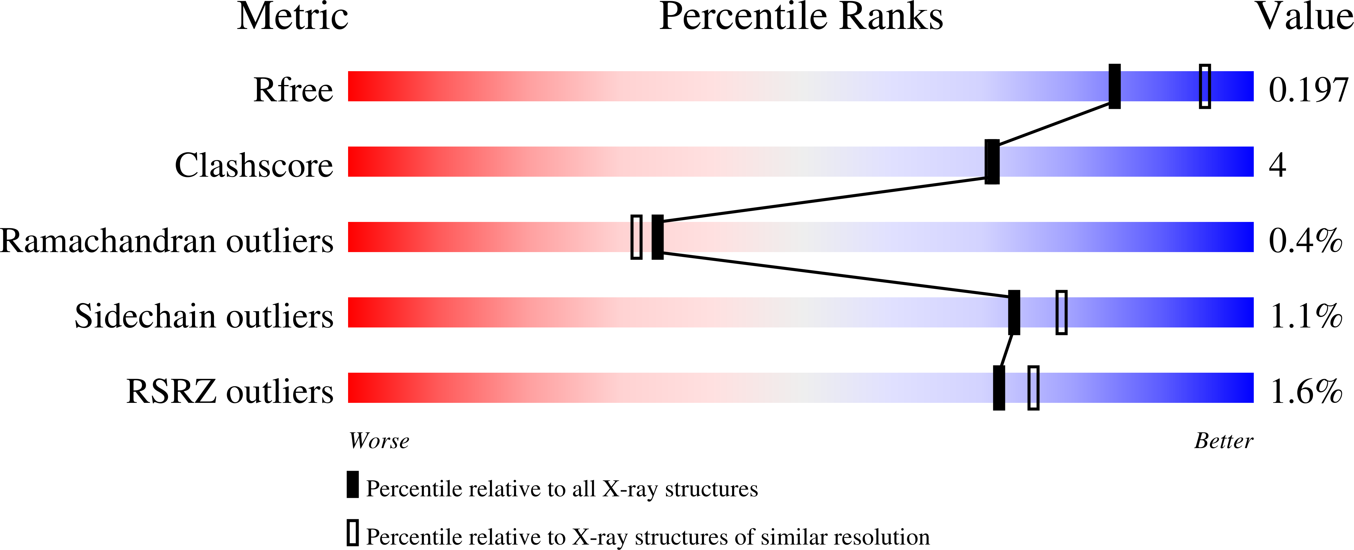
Experimental Data Snapshot
Entity ID: 1 | |||||
|---|---|---|---|---|---|
| Molecule | Chains | Sequence Length | Organism | Details | Image |
| Uridine phosphorylase | 253 | Vibrio cholerae | Mutation(s): 0 Gene Names: udp EC: 2.4.2.3 | 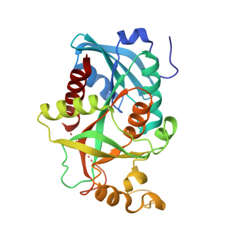 | |
UniProt | |||||
Find proteins for Q9K4U1 (Vibrio cholerae) Explore Q9K4U1 Go to UniProtKB: Q9K4U1 | |||||
Entity Groups | |||||
| Sequence Clusters | 30% Identity50% Identity70% Identity90% Identity95% Identity100% Identity | ||||
| UniProt Group | Q9K4U1 | ||||
Sequence AnnotationsExpand | |||||
| |||||
| Ligands 5 Unique | |||||
|---|---|---|---|---|---|
| ID | Chains | Name / Formula / InChI Key | 2D Diagram | 3D Interactions | |
| THM Query on THM | G [auth A] K [auth B] N [auth C] T [auth D] V [auth E] | THYMIDINE C10 H14 N2 O5 IQFYYKKMVGJFEH-XLPZGREQSA-N |  | ||
| EDO Query on EDO | O [auth C], P [auth C], Q [auth C], W [auth E] | 1,2-ETHANEDIOL C2 H6 O2 LYCAIKOWRPUZTN-UHFFFAOYSA-N |  | ||
| CL Query on CL | AA [auth F] BA [auth F] CA [auth F] H [auth A] I [auth A] | CHLORIDE ION Cl VEXZGXHMUGYJMC-UHFFFAOYSA-M |  | ||
| MG Query on MG | DA [auth F], J [auth A] | MAGNESIUM ION Mg JLVVSXFLKOJNIY-UHFFFAOYSA-N |  | ||
| NA Query on NA | M [auth B], S [auth C], Y [auth E] | SODIUM ION Na FKNQFGJONOIPTF-UHFFFAOYSA-N |  | ||
| Length ( Å ) | Angle ( ˚ ) |
|---|---|
| a = 91.796 | α = 90 |
| b = 95.905 | β = 119.96 |
| c = 91.89 | γ = 90 |
| Software Name | Purpose |
|---|---|
| SCALA | data scaling |
| PHASER | phasing |
| PHENIX | refinement |
| PDB_EXTRACT | data extraction |
| DNA | data collection |
| XSCALE | data scaling |
RCSB PDB (citation) is hosted by
RCSB PDB is a member of the

