4G3A

Crystal Structure of MAST/Orbit N-terminal domain


Experimental Data Snapshot

  • Method: X-RAY DIFFRACTION
  • Resolution: 1.99 Å
  • R-Value Free: 0.229 
  • R-Value Work: 0.188 
  • R-Value Observed: 0.190 

wwPDB Validation   3D Report Full Report


This is version 1.0 of the entry. See complete history


Literature

The structure of the TOG-like domain of Drosophila melanogaster Mast/Orbit.

De la Mora-Rey, T.Guenther, B.D.Finzel, B.C.

(2013) Acta Crystallogr Sect F Struct Biol Cryst Commun 69: 723-729

  • DOI: https://doi.org/10.1107/S1744309113015182
  • Primary Citation of Related Structures:  
    4G3A

  • PubMed Abstract: 

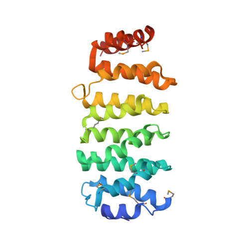
    Mast/Orbit is a nonmotor microtubule-associated protein (MAP) present in Drosophila melanogaster that reportedly binds microtubules at the plus end and is essential for mitosis. Sequence analysis has shown that the N-terminal domain (Mast-M1) resembles TOG domains from the Dis1-TOG family of proteins and stands as a representative of one of the three subclasses of divergent TOG-like domains (TOGL1) that includes human CLASP1. The crystal structure of Mast-M1 has been determined at 2.0 Å resolution and provides the first detailed structural description of any TOG-like domain. The structure confirms that Mast-M1 adopts a similar fold to the previously described Dis1-TOG domains of microtubule-binding proteins. A comparison with three known TOG-domain structures from XMAP215/Dis1 family members exposes significant differences between Mast-M1 and other TOG-domain structures in key residues at the proposed tubulin-binding edge.


  • Organizational Affiliation

    Department of Medicinal Chemistry, University of Minnesota, 308 Harvard Street SE, 8-101 Weaver-Densford Hall, Minneapolis, MN 55455, USA.


Macromolecules
Find similar proteins by:  (by identity cutoff)  |  3D Structure
Entity ID: 1
MoleculeChains Sequence LengthOrganismDetailsImage
CLIP-associating protein
A, B
237Drosophila melanogasterMutation(s): 0 
Gene Names: CG32435chbCLASPMastMESR7orbit
UniProt
Find proteins for Q9NBD7 (Drosophila melanogaster)
Explore Q9NBD7 
Go to UniProtKB:  Q9NBD7
Entity Groups  
Sequence Clusters30% Identity50% Identity70% Identity90% Identity95% Identity100% Identity
UniProt GroupQ9NBD7
Sequence Annotations
Expand
  • Reference Sequence
Small Molecules
Modified Residues  1 Unique
IDChains TypeFormula2D DiagramParent
MSE
Query on MSE
A, B
L-PEPTIDE LINKINGC5 H11 N O2 SeMET
Experimental Data & Validation

Experimental Data

  • Method: X-RAY DIFFRACTION
  • Resolution: 1.99 Å
  • R-Value Free: 0.229 
  • R-Value Work: 0.188 
  • R-Value Observed: 0.190 
  • Space Group: P 21 21 21
Unit Cell:
Length ( Å )Angle ( ˚ )
a = 36.18α = 90
b = 97.579β = 90
c = 149.346γ = 90
Software Package:
Software NamePurpose
PHENIXrefinement

Structure Validation

View Full Validation Report


Entry History 

Deposition Data

Revision History  (Full details and data files)

  • Version 1.0: 2013-07-17
    Type: Initial release