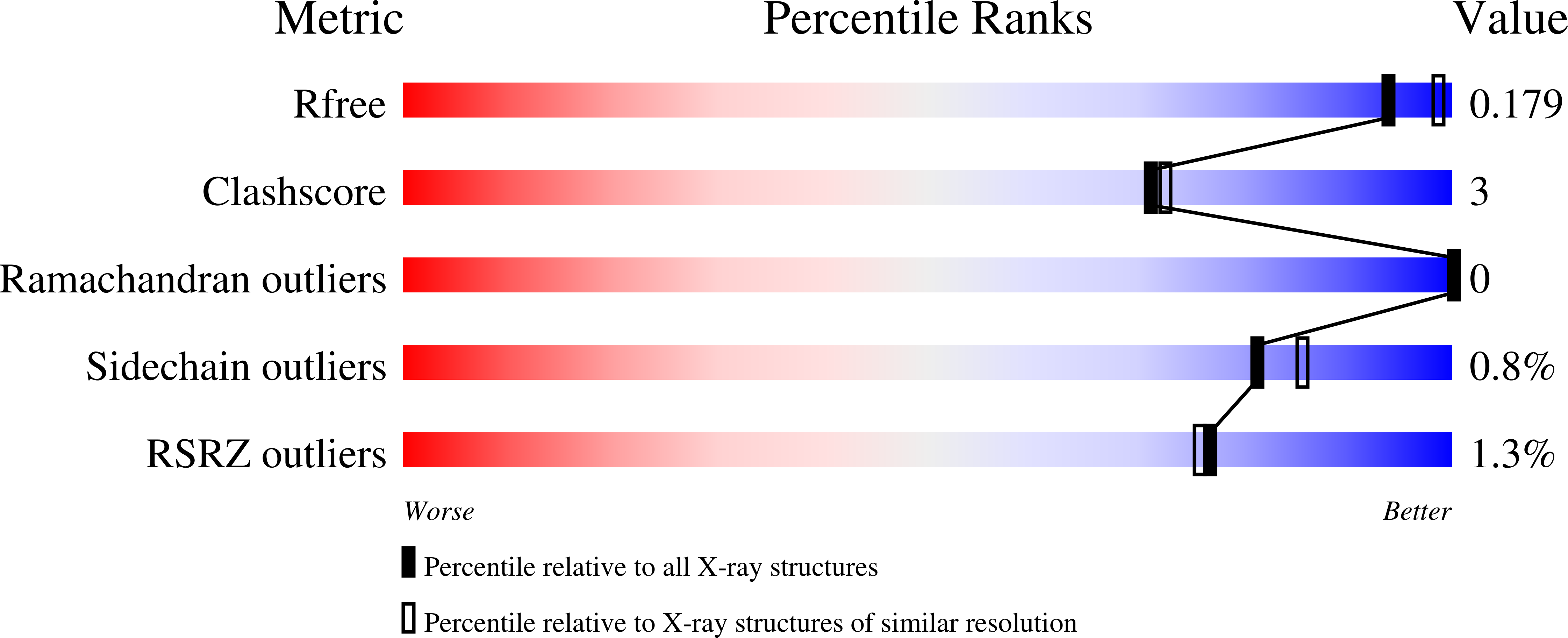
4FWU

Crystal structure of glutaminyl cyclase from drosophila melanogaster in space group I4


Experimental Data Snapshot

  • Method: X-RAY DIFFRACTION
  • Resolution: 2.00 Å
  • R-Value Free: 0.208 
  • R-Value Work: 0.166 
  • R-Value Observed: 0.170 

wwPDB Validation   3D Report Full Report


This is version 2.1 of the entry. See complete history


Literature

Structure of glutaminyl cyclase from Drosophila melanogaster in space group I4.

Kolenko, P.Koch, B.Rahfeld, J.U.Schilling, S.Demuth, H.U.Stubbs, M.T.

(2013) Acta Crystallogr Sect F Struct Biol Cryst Commun 69: 358-361

  • DOI: https://doi.org/10.1107/S1744309113005575
  • Primary Citation of Related Structures:  
    4FWU

  • PubMed Abstract: 

    The structure of ligand-free glutaminyl cyclase (QC) from Drosophila melanogaster (DmQC) has been determined in a novel crystal form. The protein crystallized in space group I4, with unit-cell parameters a = b = 122.3, c = 72.7 Å. The crystal diffracted to a resolution of 2 Å at the home source. The structure was solved by molecular replacement and was refined to an R factor of 0.169. DmQC exhibits a typical α/β-hydrolase fold. The electron density of three monosaccharides could be localized. The accessibility of the active site will facilitate structural studies of novel inhibitor-binding modes.


  • Organizational Affiliation

    Department of Physical Biochemistry, Institute of Biochemistry and Biotechnology, MLU, Kurt-Mothes-Strasse 3, 06120 Halle, Germany. petr.kolenko@gmail.com


Macromolecules
Find similar proteins by:  (by identity cutoff)  |  3D Structure
Entity ID: 1
MoleculeChains Sequence LengthOrganismDetailsImage
CG32412312Drosophila melanogasterMutation(s): 0 
Gene Names: QCCG10487CG32412Dmel_CG32412
EC: 2.3.2.5 (PDB Primary Data), 3.4 (PDB Primary Data)
UniProt
Find proteins for Q9VRQ9 (Drosophila melanogaster)
Explore Q9VRQ9 
Go to UniProtKB:  Q9VRQ9
Entity Groups  
Sequence Clusters30% Identity50% Identity70% Identity90% Identity95% Identity100% Identity
UniProt GroupQ9VRQ9
Sequence Annotations
Expand
  • Reference Sequence
Oligosaccharides

Help

Entity ID: 2
MoleculeChains Length2D Diagram Glycosylation3D Interactions
beta-D-mannopyranose-(1-4)-2-acetamido-2-deoxy-beta-D-glucopyranose-(1-4)-2-acetamido-2-deoxy-beta-D-glucopyranose
B
3N-Glycosylation
Glycosylation Resources
GlyTouCan:  G15407YE
GlyCosmos:  G15407YE
GlyGen:  G15407YE
Small Molecules
Ligands 3 Unique
IDChains Name / Formula / InChI Key2D Diagram3D Interactions
SO4
Query on SO4

Download Ideal Coordinates CCD File 
F [auth A],
G [auth A],
H [auth A],
J [auth A],
K [auth A]
SULFATE ION
O4 S
QAOWNCQODCNURD-UHFFFAOYSA-L
ZN
Query on ZN

Download Ideal Coordinates CCD File 
C [auth A]ZINC ION
Zn
PTFCDOFLOPIGGS-UHFFFAOYSA-N
EDO
Query on EDO

Download Ideal Coordinates CCD File 
D [auth A],
E [auth A],
I [auth A]
1,2-ETHANEDIOL
C2 H6 O2
LYCAIKOWRPUZTN-UHFFFAOYSA-N
Experimental Data & Validation

Experimental Data

  • Method: X-RAY DIFFRACTION
  • Resolution: 2.00 Å
  • R-Value Free: 0.208 
  • R-Value Work: 0.166 
  • R-Value Observed: 0.170 
  • Space Group: I 4
Unit Cell:
Length ( Å )Angle ( ˚ )
a = 122.344α = 90
b = 122.344β = 90
c = 72.736γ = 90
Software Package:
Software NamePurpose
CrystalCleardata collection
MOLREPphasing
REFMACrefinement
XDSdata reduction
SCALAdata scaling

Structure Validation

View Full Validation Report


Entry History 

Deposition Data

Revision History  (Full details and data files)

  • Version 1.0: 2012-09-05
    Type: Initial release
  • Version 1.1: 2013-04-10
    Changes: Database references
  • Version 1.2: 2013-04-17
    Changes: Database references
  • Version 2.0: 2020-07-29
    Type: Remediation
    Reason: Carbohydrate remediation
    Changes: Atomic model, Data collection, Derived calculations, Structure summary
  • Version 2.1: 2023-09-13
    Changes: Data collection, Database references, Refinement description, Structure summary